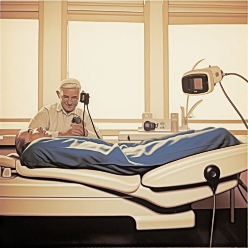
Medical Imaging Techniques
Introduction
Medical imaging is a discipline within the field of medical science that utilizes various techniques and processes to create visual representations of the interior of the body for clinical analysis and medical intervention. Medical imaging seeks to reveal internal structures hidden by the skin and bones, as well as to diagnose and treat disease. It also establishes a database of normal anatomy and physiology to make it possible to identify abnormalities.
History
The history of medical imaging dates back to the discovery of X-rays by Wilhelm Conrad Roentgen in 1895. This discovery revolutionized the field of medicine by providing a non-invasive means to examine the internal structures of the body. Over the years, numerous other imaging techniques have been developed, each with its unique advantages and applications.
Types of Medical Imaging Techniques
There are several types of medical imaging techniques, each used for different purposes and providing different types of information.
Radiography
Radiography is the use of X-rays to view a single plane of the body in a two-dimensional image. It is the oldest and most frequently used form of medical imaging. Radiography is primarily used for identifying and diagnosing bone fractures, certain infections, and locating foreign objects in the body.
Magnetic Resonance Imaging (MRI)
MRI uses a magnetic field and radio waves to create detailed images of the organs and tissues within the body. Unlike X-rays and CT scans, MRI does not use ionizing radiation. It is often used for disease detection, diagnosis, and treatment monitoring.
Computed Tomography (CT)
CT, also known as a CAT scan, uses a combination of X-rays and a computer to create images of the body. It can create both two-dimensional and three-dimensional images of the body, allowing for detailed examination of the body's structures.
Ultrasound
Ultrasound imaging, also known as sonography, uses high-frequency sound waves to visualize soft tissues such as muscles, tendons, and internal organs. Because ultrasound images are captured in real-time, they can show movement of the body's internal organs as well as blood flowing through blood vessels.
Positron Emission Tomography (PET)
PET scans use a radioactive substance, known as a tracer, to look for disease in the body. PET scans can measure important body functions, such as blood flow, oxygen use, and glucose metabolism, to help doctors evaluate how well organs and tissues are functioning.
Applications
Medical imaging techniques are used in various medical fields for different purposes. They are used in radiology, cardiology, oncology, neurology, and many other fields. They are used for diagnosing diseases, planning treatments, guiding medical procedures, and monitoring the progress of diseases.
Future Developments
The future of medical imaging lies in the development of new technologies and the improvement of existing ones. This includes the development of more advanced imaging techniques, such as molecular imaging, which allows for the visualization of cellular and molecular processes in the body. Additionally, advances in artificial intelligence and machine learning are expected to play a significant role in the future of medical imaging.
See Also


