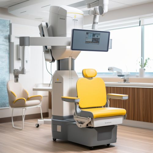
Radiography
Introduction
Radiography is a type of medical imaging technology that utilizes X-rays to visualize the internal structures of the body. It is a vital tool in the diagnosis and treatment of various medical conditions, and is widely used in hospitals and clinics around the world.
History
Radiography was first discovered in 1895 by Wilhelm Conrad Roentgen, a German physicist. Roentgen discovered that a cathode ray tube could produce a new kind of ray, which he called "X-rays". These rays were capable of passing through most substances, including human tissue, but were blocked by denser materials such as bone and metal. This discovery revolutionized the field of medicine, as it allowed doctors to see inside the human body without the need for invasive procedures.
Principles of Radiography
Radiography works by passing a controlled amount of X-ray radiation through the body. The X-rays are absorbed by different tissues to varying degrees, depending on their density and composition. Denser tissues, such as bones, absorb more X-rays and appear white on the radiograph, while less dense tissues, such as muscles and organs, absorb fewer X-rays and appear darker. This contrast allows the radiographer to distinguish between different types of tissue and identify any abnormalities.
Types of Radiography
There are several types of radiography, each with its own specific uses and advantages.
Plain Radiography
Plain radiography, also known as conventional radiography, is the most common type of radiography. It is often used to diagnose fractures, infections, and tumors in the bones and lungs.
Fluoroscopy
Fluoroscopy is a type of radiography that uses a continuous or pulsed X-ray beam to create real-time images of the body. This allows doctors to observe the movement of internal structures, such as the heart and digestive system, and perform procedures such as angiograms and barium enemas.
Computed Tomography
Computed Tomography (CT) is a type of radiography that uses a rotating X-ray machine to create detailed, cross-sectional images of the body. CT scans provide more detailed information than conventional radiographs and are often used to diagnose and monitor conditions such as cancer, heart disease, and stroke.
Safety and Risks
While radiography is a valuable diagnostic tool, it does involve exposure to ionizing radiation, which can potentially cause harm. However, the benefits of radiography typically outweigh the risks, and precautions are taken to minimize exposure. These include using the lowest possible dose of radiation, shielding sensitive parts of the body, and avoiding unnecessary repeat examinations.
Future Developments
Advancements in technology continue to improve the capabilities of radiography. Digital radiography, which uses digital detectors instead of traditional film, offers several advantages, including better image quality, lower radiation doses, and the ability to manipulate and enhance images. Other areas of research include the development of new contrast agents and techniques to improve image quality and the use of artificial intelligence to assist in image interpretation.


