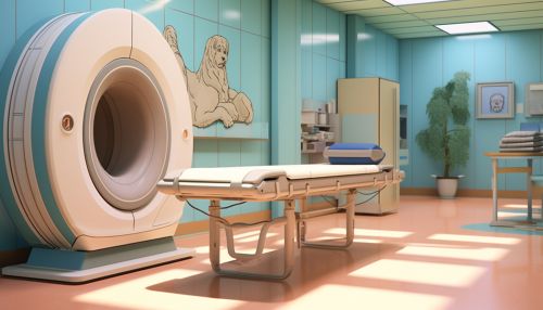
Positron Emission Tomography (PET)
Introduction
Positron Emission Tomography (PET) is a type of nuclear medicine imaging technique that produces a three-dimensional image or picture of functional processes in the body. The system detects pairs of gamma rays emitted indirectly by a positron-emitting radionuclide (tracer), which is introduced into the body on a biologically active molecule. Images of tracer concentration in 3-dimensional space within the body are then constructed by computer analysis.
History
The concept of PET was first proposed by James Robertson and his student Yale University student Gordon Brownell in 1951. The first PET scanner was built in 1961 by James Robertson and his team at Brookhaven National Laboratory. The first commercial PET scanners were produced in the 1970s and were primarily used for research purposes. The use of PET for medical diagnosis did not become common until the 1980s.
Principle of Operation
PET is a type of imaging that uses a radioactive tracer to visualize the body's function and metabolism, rather than its structure. The tracer is a biologically active molecule that has been labeled with a positron-emitting radionuclide. When the tracer is introduced into the body, it accumulates in the target tissue or organ. The radionuclide then decays, emitting a positron. When the positron encounters an electron, it undergoes annihilation, resulting in the emission of two gamma rays in opposite directions. These gamma rays are detected by the PET scanner, which uses this information to construct a three-dimensional image of the tracer distribution within the body.
Tracers
The choice of tracer depends on the process that is to be imaged. The most common tracer is fluorodeoxyglucose (FDG), a glucose analog that is taken up by cells and phosphorylated by hexokinase, whose mitochondrial form is significantly upregulated in rapidly growing cancers. Other tracers are used to image the tissue concentration of other types of molecules.
Image Reconstruction
The data collected by the PET scanner is a list of 'coincidence events' representing near-simultaneous detection of annihilation photons by a pair of detectors. These data are then sorted by the detected angle of coincidence and, using a form of tomographic reconstruction, an image of the distribution of radionuclides within the patient is created.


Clinical Applications
PET imaging is primarily used in clinical oncology, with FDG-PET scans being the most common. These scans are used to detect, diagnose, and monitor the treatment of various types of cancers. PET can also be used in neurology and cardiology to diagnose and monitor diseases such as Alzheimer's, Parkinson's, and coronary artery disease.
Advantages and Limitations
One of the main advantages of PET is its ability to measure metabolic activity and physiological function, which can be more important than anatomy in determining the presence and severity of disease. However, PET has several limitations, including a relatively low spatial resolution and the need for a cyclotron on site or nearby to produce the short-lived radionuclides for PET scanning.
Future Developments
The future of PET imaging lies in the development of new tracers that can target specific types of cells or processes. This will allow for more precise imaging of disease processes and may also enable new applications of PET imaging in areas such as drug development and gene therapy.
