Computed Tomography (CT)
Introduction
Computed Tomography (CT) is a diagnostic imaging technique that uses computer-processed combinations of many X-ray measurements taken from different angles to produce cross-sectional (tomographic) images (virtual 'slices') of specific areas of a scanned object, allowing the user to see inside the object without cutting.


History
The development of CT started with the invention of the X-ray by Wilhelm Conrad Roentgen in 1895. However, it wasn't until the 1970s that the first commercially viable CT scanner was developed by Godfrey Hounsfield at EMI Laboratories in England. This early machine, known as the EMI-Scanner, was capable of producing a single slice of the brain in about 5 minutes. The first clinical CT scanners were installed between 1974 and 1976.
Principles of Operation
CT operates by using an X-ray generator that rotates around the patient, shooting an individual thin X-ray beam through the patient to a detector located on the opposite side. The X-ray beam intensity is measured and the data is sent to a computer for processing. The computer then uses a mathematical algorithm to reconstruct an image of the section that was scanned. The resulting image is a two-dimensional slice of the patient's body.
CT Scanning Procedure
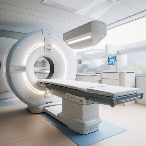
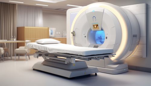
During a CT scan, the patient lies on a table that moves through a circular opening in the CT machine. The X-ray tube and detectors within the machine rotate around the patient, capturing images from multiple angles. These images are then processed by a computer to create cross-sectional images of the body.
Applications
CT scans are used in a variety of medical applications, including:
- Diagnostic imaging: CT is often used to diagnose diseases and conditions such as cancer, cardiovascular disease, infectious disease, appendicitis, and trauma. - Interventional procedures: CT can guide biopsies and other procedures such as abscess drainages and minimally invasive tumor treatments. - Radiation therapy planning: CT is often used in planning radiation therapy, as it allows doctors to determine the exact location, shape, and size of the tumor to be treated.
Risks and Benefits
Like all medical procedures, CT scans have both risks and benefits. The main risk associated with CT scans is exposure to ionizing radiation, which can increase the risk of cancer. However, the benefits of accurate diagnosis and treatment planning often outweigh these risks.
Future Developments
Future developments in CT technology include spectral (or dual-energy) CT, which uses two different X-ray energy levels to produce images, and 4D CT, which adds the dimension of time to the 3D images produced by CT.
See Also
- Magnetic Resonance Imaging (MRI) - Positron Emission Tomography (PET) - Ultrasound Imaging
