Magnetic Resonance Imaging (MRI)
Introduction
Magnetic Resonance Imaging (MRI) is a non-invasive imaging technology that produces three dimensional detailed anatomical images. It is often used for disease detection, diagnosis, and treatment monitoring. It is based on sophisticated technology that excites and detects the change in direction of the rotational axis of protons found in the water that makes up living tissues[1].
Physics of MRI
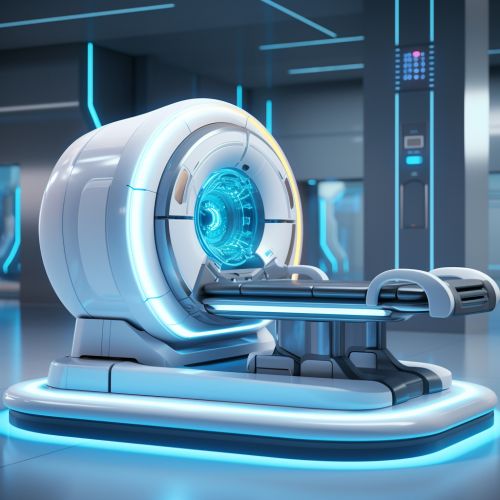
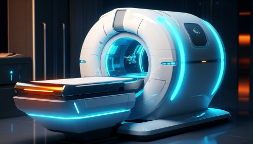
MRI is a medical imaging technique used in radiology to visualize internal structures of the body in detail. MRI makes use of the property of nuclear magnetic resonance (NMR) to image nuclei of atoms inside the body. An MRI scanner is a device in which the patient lies within a large, powerful magnet where the magnetic field is used to align the magnetization of some atomic nuclei in the body, and radio frequency fields are used to systematically alter the alignment of this magnetization. This causes the nuclei to produce a rotating magnetic field detectable by the scanner—and this information is recorded to construct an image of the scanned area of the body[2].
Components of an MRI system
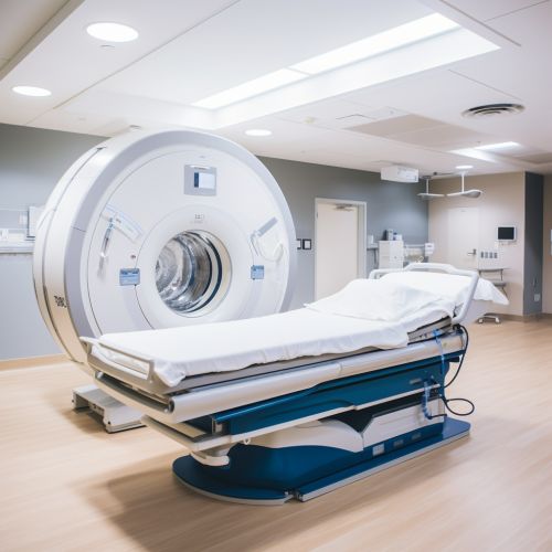
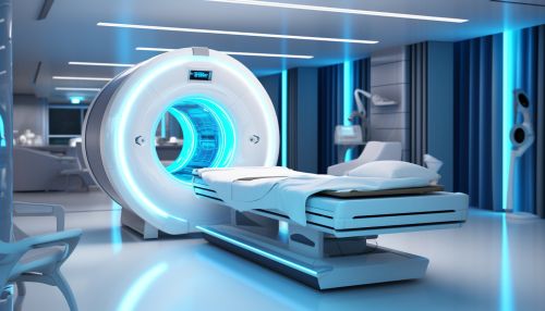
An MRI system consists of a large, heavy magnet, radio frequency (RF) coils, gradient coils, and a computer system. The magnet is the largest and most expensive component of the system, and its strength is measured in teslas. Most clinical MRI systems are superconducting magnets, which means that they maintain a high magnetic field with no power input, thus making them very efficient[3].
MRI Procedure
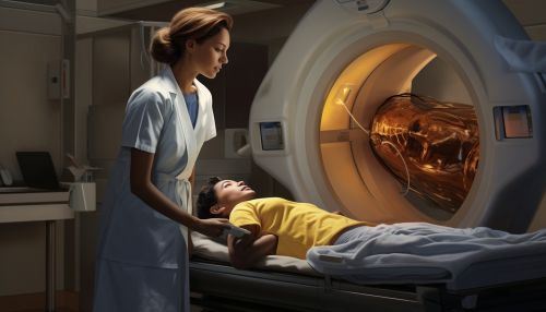
The patient is placed on a moveable bed that is inserted into the magnet. The patient is moved into the magnet until the part of the body to be scanned is in the center of the magnet. The RF coil is placed around the part of the body to be imaged. The RF coil is a small antenna that is capable of transmitting and receiving radio waves. Once the patient is properly positioned, the scan can begin[4].
Applications of MRI
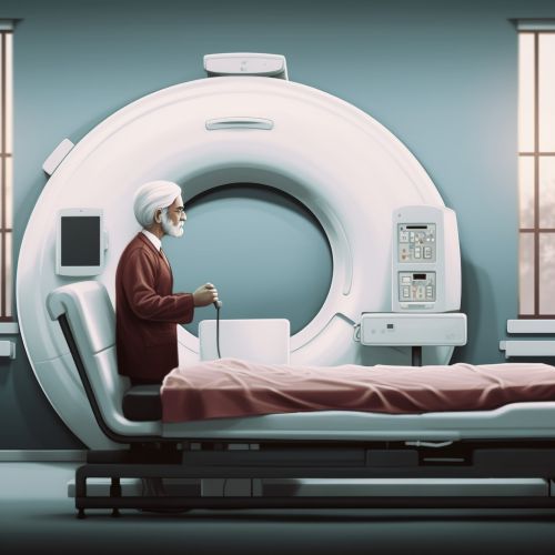
MRI has a wide range of applications in medical diagnosis and more than 25,000 scanners are estimated to be in use worldwide. MRI affects the body in a very different way than X-ray or CT scanning, and is considered to have no known harmful effects when used appropriately. It is used for a wide range of applications, including imaging of soft tissues such as the brain, spinal cord, muscles, and heart[5].
Safety and contraindications
Because MRI uses strong magnets, the presence of metal in your body can be a safety hazard if attracted to the magnet. Even if not attracted to the magnet, metal objects can distort the MRI image. Therefore, metal objects are carefully screened for prior to an MRI scan. Patients with certain implants, cochlear implants, certain types of vascular clips, or metal fragments in their eyes are advised not to have an MRI scan[6].


Advantages and disadvantages
MRI has many advantages over other imaging techniques. It can image in any plane, has no known harmful effects, and is capable of high resolution imaging. The main disadvantage of MRI is its high cost, and the fact that it cannot be used in the presence of certain implants or medical devices, such as pacemakers[7].
Future of MRI
The future of MRI is very promising, with new techniques and applications constantly being developed. These include functional MRI (fMRI), which can be used to measure brain activity, and diffusion tensor imaging (DTI), which can be used to image nerve pathways in the brain[8].
See Also
References
[1] "Magnetic Resonance Imaging (MRI)". National Institute of Biomedical Imaging and Bioengineering. [2] "MRI: A Guided Tour". Magnetic Resonance Imaging. McGraw Hill. [3] "Introduction to MRI". University of Nottingham. [4] "MRI Procedure". Johns Hopkins Medicine. [5] "Applications of MRI". RadiologyInfo.org. [6] "MRI Safety". American College of Radiology. [7] "Advantages and Disadvantages of MRI". RadiologyInfo.org. [8] "The Future of MRI". Radiology Today.
