Computed Tomography
Introduction
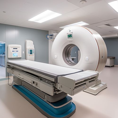
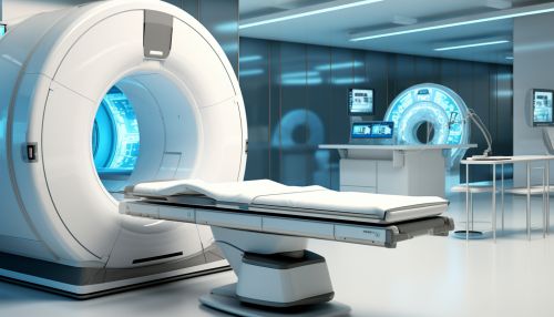
Computed Tomography (CT) is a type of medical imaging technique that uses computer-processed combinations of many X-ray measurements taken from different angles to produce cross-sectional (tomographic) images (virtual 'slices') of specific areas of a scanned object. These images can be manipulated in various planes and also can be observed as three-dimensional images.
History
The concept of CT was first developed in 1967 by the British engineer Godfrey N. Hounsfield. His work, which involved the construction of a prototype CT machine, was later supported by the British company Electric and Musical Industries Ltd. The first clinical CT scanners were installed between 1974 and 1976. The original systems were dedicated to head imaging only, but whole body systems were introduced in 1976.
Principles of Operation
CT operates by using an X-ray generator that rotates around the object; X-ray detectors are positioned on the opposite side of the circle from the X-ray source. A fan-shaped beam of X-rays is created as the rotating frame carrying the X-ray tube and detectors passes around the patient. The attenuation of the X-rays is measured from different angles, providing multiple measurements from different angles.


Image Reconstruction
The data obtained from the measurements is then used to reconstruct a three-dimensional image of the inside of the object. This process is carried out by a computer using a technique known as filtered back projection. The resulting image is a high-resolution 3D representation of the scanned object.
Clinical Applications
CT is used in medicine as a diagnostic tool and as a guide for interventional procedures. It is also used in other fields such as nondestructive materials testing. In healthcare, CT is used to diagnose disease, guide biopsies and other procedures, plan surgery, and monitor therapy.
Advantages and Limitations
CT scanning has several advantages over traditional radiographic techniques. It can image a combination of soft tissue, bone, and blood vessels all at the same time. It also provides more detailed information than conventional X-ray imaging. However, CT scans have limitations. They expose the patient to more radiation than regular X-rays, and they can't be used to image certain areas of the body, such as the lungs and bones.
Future Developments
Future developments in CT technology include reducing radiation exposure, increasing image quality, and improving machine learning algorithms for image reconstruction and analysis.
