Fluoroscopy
Introduction
Fluoroscopy is a type of medical imaging that uses X-rays to obtain real-time moving images of the interior of an object. It is used in both diagnostic and therapeutic procedures to visualize the movement of a body organ, a device or contrast agent through the body.
History
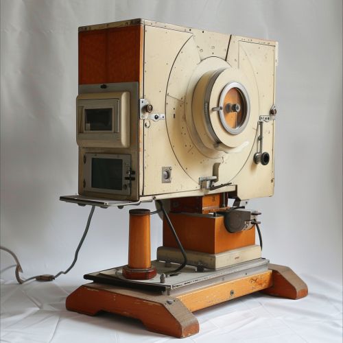
The discovery of X-rays by Röntgen in 1895 led to the development of fluoroscopy later that year by American surgeon, Edison. The first fluoroscopes consisted simply of an X-ray source and a fluorescent screen, between which the patient was placed.


Principles
Fluoroscopy works on the principle of differential absorption of X-ray photons by different tissues. The X-rays pass through the patient and are absorbed by the detector, which then converts the X-rays into a visible image. The image is then displayed on a monitor for real-time viewing.
Procedure
During a fluoroscopy procedure, the patient is positioned between the X-ray source and the detector. The X-ray tube emits a beam of X-rays which pass through the patient's body and are absorbed by the detector. The detector then converts the X-rays into an electrical signal which is processed to produce a visible image on a monitor.
Applications
Fluoroscopy is used in a wide range of medical procedures, including angiography, barium studies, cardiac catheterization, orthopedic surgery, and urological procedures. It is also used in non-medical applications such as in airport security scanners.
Risks and Safety
While fluoroscopy provides valuable diagnostic and therapeutic information, it also exposes the patient to ionizing radiation, which can potentially cause harm. Therefore, it is important to use the lowest possible dose of radiation and to limit the duration of the procedure. Protective measures such as lead aprons and thyroid shields are also used to minimize radiation exposure.
Future Developments
Advancements in technology are continually improving the quality and safety of fluoroscopy. These include the development of digital detectors, which provide higher image quality and lower radiation doses, and the use of computer algorithms to enhance image quality and reduce noise.
