Medical imaging
Overview
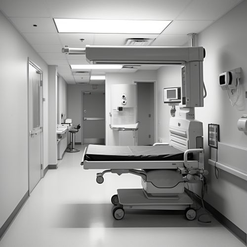
Medical imaging is a discipline within the field of radiology that utilizes various techniques to visualize the interior of the human body for diagnostic and therapeutic purposes. The images produced can be used to examine the function of internal organs, identify disease or injury, and guide medical procedures. Medical imaging has revolutionized healthcare, allowing for non-invasive diagnosis and treatment of a wide range of conditions.
History
The history of medical imaging can be traced back to the discovery of radiography by Wilhelm Conrad Roentgen in 1895. This marked the beginning of a new era in medicine, as it allowed doctors to see inside the human body without surgery. Over the years, many other imaging techniques have been developed, including ultrasound, computed tomography (CT), magnetic resonance imaging (MRI), and positron emission tomography (PET).
Types of Medical Imaging
There are several types of medical imaging, each with its own advantages and limitations. The choice of imaging technique depends on the patient's symptoms, the part of the body being examined, and the clinical question being asked.
Radiography
Radiography is the oldest and most commonly used form of medical imaging. It uses X-rays to produce images of the body's internal structures. Radiography is particularly useful for imaging bones and detecting abnormalities in the lungs.
Ultrasound
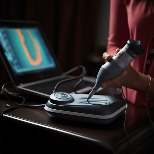
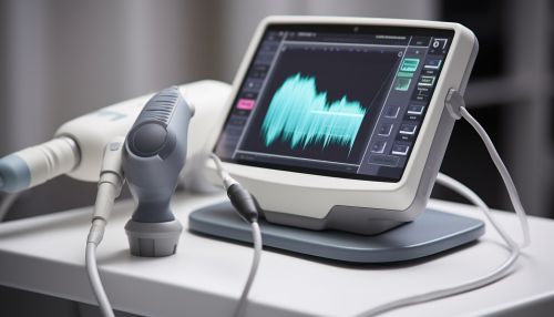
Ultrasound uses high-frequency sound waves to create images of the body's internal organs. It is a safe and non-invasive method of imaging, often used in obstetrics to monitor the development of a fetus during pregnancy.
Computed Tomography (CT)
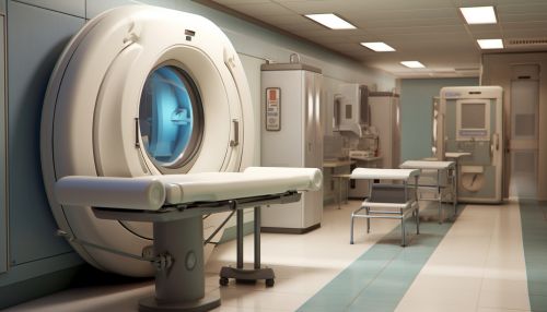
CT scanning combines X-ray technology with computer processing to produce detailed, cross-sectional images of the body. It is often used to diagnose conditions such as cancer, cardiovascular disease, and trauma.
Magnetic Resonance Imaging (MRI)
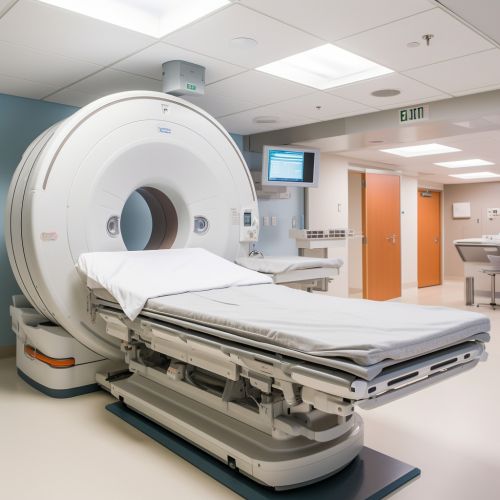
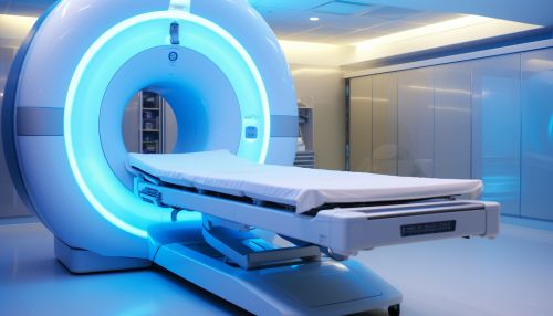
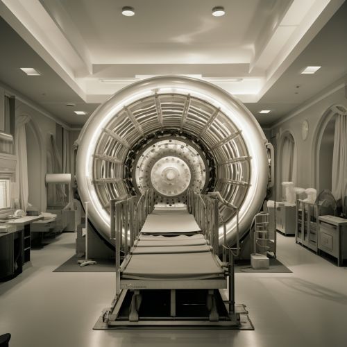
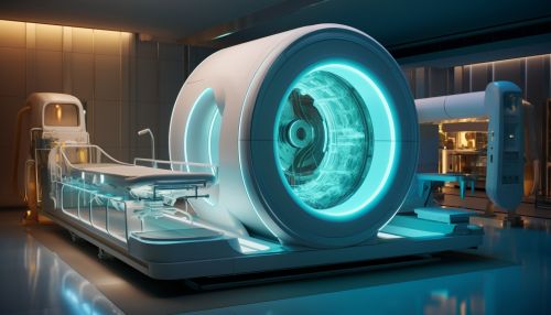
MRI uses a powerful magnetic field and radio waves to generate detailed images of the body's internal structures. It is particularly useful for imaging soft tissues and organs such as the brain, spinal cord, and heart.
Positron Emission Tomography (PET)
PET uses a radioactive tracer to visualize metabolic processes in the body. It is often used in conjunction with CT or MRI to provide detailed information about both the structure and function of tissues and organs.
Applications
Medical imaging is used in a wide range of clinical applications, from diagnosis and treatment planning to monitoring disease progression and guiding surgical procedures.
Diagnosis
Medical imaging plays a crucial role in the diagnosis of many conditions. It can reveal abnormalities in the body's structure or function that may indicate disease, such as tumors, fractures, or blockages in blood vessels.
Treatment Planning
Imaging can also be used to plan treatments, such as surgery or radiation therapy. For example, CT or MRI scans can be used to determine the exact location and size of a tumor, which can help doctors plan the most effective treatment.
Monitoring Disease Progression
Medical imaging can be used to monitor the progression of a disease over time. This can help doctors assess the effectiveness of treatment and make necessary adjustments.
Guiding Procedures
Imaging techniques such as ultrasound or fluoroscopy can be used to guide medical procedures. For example, doctors may use imaging to guide the placement of a catheter or to perform a biopsy.
Future Developments
The field of medical imaging continues to evolve, with ongoing research aimed at improving image quality, reducing radiation exposure, and developing new imaging techniques. Advances in artificial intelligence and machine learning are also expected to play a significant role in the future of medical imaging, with potential applications in image interpretation, diagnosis, and treatment planning.
