Neuroradiology
Overview
Neuroradiology is a subspecialty of radiology focusing on the diagnosis and characterization of abnormalities of the central nervous system, spine, and head and neck using neuroimaging techniques. These techniques include Computed Tomography (CT), Magnetic Resonance Imaging (MRI), and Ultrasound (US).


History
The field of neuroradiology began to emerge in the early 20th century with the advent of x-ray technology. The first neuroradiological procedure, a cerebral angiogram, was performed in 1927 by Portuguese neurologist Egas Moniz. The development of non-invasive imaging techniques such as CT and MRI in the late 20th century greatly expanded the capabilities of neuroradiology.
Techniques
Neuroradiology employs a variety of imaging techniques to diagnose and treat diseases of the nervous system. These include:
Computed Tomography
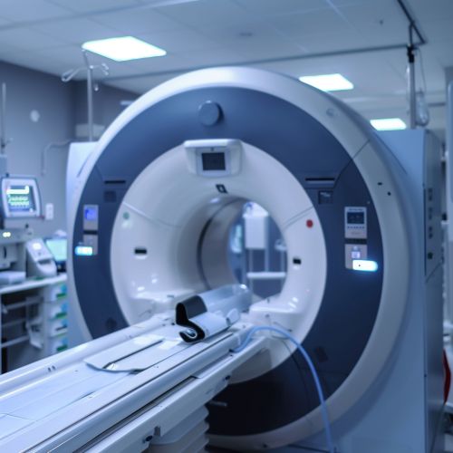
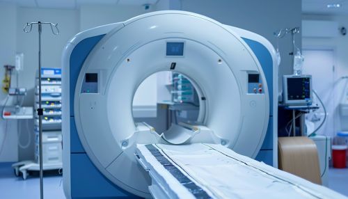
Computed Tomography (CT) uses x-rays to produce cross-sectional images of the body. In neuroradiology, CT can be used to quickly visualize the brain and spine, and is particularly useful in acute situations such as stroke or traumatic brain injury.
Magnetic Resonance Imaging
Magnetic Resonance Imaging (MRI) uses magnetic fields and radio waves to generate detailed images of the body's soft tissues. MRI is often used in neuroradiology to visualize the brain and spinal cord, and can provide more detailed information than CT for many types of neurological conditions.
Ultrasound
Ultrasound (US) uses high-frequency sound waves to create images of the body's internal structures. In neuroradiology, US is often used to image the blood vessels of the brain and neck, as well as the spinal cord in infants.
Applications
Neuroradiology plays a crucial role in the diagnosis and treatment of a wide range of neurological conditions. These include:
Stroke
In cases of suspected stroke, neuroradiological techniques such as CT and MRI can be used to quickly visualize the brain and determine the type and extent of the stroke. This information is critical in guiding treatment decisions.
Brain Tumors
Neuroradiology is essential in the diagnosis and management of brain tumors. Imaging techniques such as MRI can provide detailed information about the size, location, and characteristics of the tumor, which can guide surgical planning and monitor the effectiveness of treatment.
Spinal Disorders
Neuroradiology is also used to diagnose and treat a variety of spinal disorders, including herniated discs, spinal stenosis, and spinal cord injuries. Imaging techniques such as CT and MRI can provide detailed images of the spine, helping to guide treatment decisions.
Future Directions
The field of neuroradiology continues to evolve with advances in imaging technology. New techniques such as functional MRI (fMRI), which allows for the visualization of brain activity, and diffusion tensor imaging (DTI), which can map the brain's white matter tracts, are expanding the capabilities of neuroradiology. These advances promise to improve our understanding of the brain and spinal cord, and to enhance the diagnosis and treatment of neurological conditions.
