Magnetic Resonance Imaging
Introduction
Magnetic Resonance Imaging (MRI) is a non-invasive medical imaging technique used in radiology to visualize detailed internal structures of the body. MRI scanners use strong magnetic fields, magnetic field gradients, and radio waves to generate images of the organs in the body. MRI does not involve X-rays or the use of ionizing radiation, which distinguishes it from CT or CAT scans.
History
MRI technology was first developed in the 1970s with significant contributions from scientists such as Raymond Damadian and Paul Lauterbur. Damadian was the first to perform a full body scan of a human being in 1977 to detect cancer. Lauterbur developed the idea of using gradient fields, which allowed for clearer and more precise images to be produced.


Physics of MRI
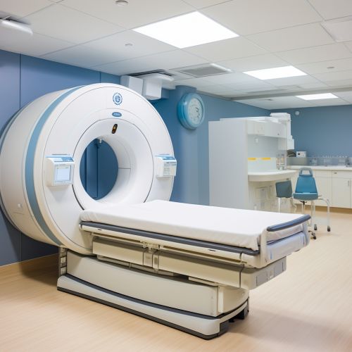
MRI uses a large magnetic field and radio waves to generate images of the body. The patient is placed inside a large cylinder containing the magnet. This magnetic field aligns the protons in the body, and a radio frequency current is then applied, causing the protons to alter their alignment relative to the field. When the field is turned off, the protons return to their normal alignment, and in doing so, they produce a signal that can be measured and used to produce an image of the body.
MRI Machine Components
An MRI scanner consists of several key components, including the main magnet, gradient system, RF system, and computer system.
- The main magnet creates a strong magnetic field around the patient. This magnet is typically superconducting, meaning it has no electrical resistance and can maintain a high magnetic field indefinitely without power input.
- The gradient system is used to vary the magnetic field across the patient's body, which allows different slices of the body to be imaged.
- The RF system generates the radio frequency pulses that are used to excite the protons in the body and create the MRI signal.
- The computer system controls the operation of the other components and processes the MRI signal to create the final image.
MRI Techniques
There are several different techniques used in MRI, each with its own advantages and disadvantages. These include T1-weighted imaging, T2-weighted imaging, diffusion-weighted imaging (DWI), and functional MRI (fMRI).
- T1-weighted imaging is useful for visualizing fat, producing images where fat appears bright. It is often used in brain imaging.
- T2-weighted imaging is useful for detecting edema and inflammation. In T2 images, water and fluid appear bright.
- Diffusion-weighted imaging (DWI) is used to measure the random Brownian motion of water molecules within a voxel of tissue.
- Functional MRI (fMRI) is used to determine the specific brain areas associated with a particular mental process. It measures changes in blood flow related to neural activity.
Clinical Applications
MRI has a wide range of clinical applications, including neuroimaging, cardiovascular imaging, musculoskeletal imaging, liver and gastrointestinal imaging, and oncological imaging. It is particularly useful for imaging soft tissues and organs like the brain, spinal cord, muscles, and heart.
Safety and Contraindications
While MRI is generally considered safe, there are some contraindications. Patients with certain types of metallic implants, pacemakers, cochlear implants, certain types of vascular clips, or metal fragments in their eyes may not be suitable for MRI. Additionally, the loud noise produced by the machine can be distressing to some patients, and the confined space can cause claustrophobia.
Future Developments
Future developments in MRI technology include the development of faster scanning techniques, higher resolution imaging, functional imaging and molecular imaging. The goal of these developments is to increase the diagnostic accuracy, safety, and patient comfort of MRI scans.
