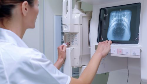
Diagnostic radiography
Introduction
Diagnostic radiography is a branch of medical imaging that uses radiation to view the internal structures of the body. It is a non-invasive procedure that allows medical professionals to diagnose and monitor various medical conditions, diseases, and injuries. Diagnostic radiography plays a crucial role in modern medicine, providing valuable information that aids in the effective treatment of patients.
History
The discovery of X-rays by Röntgen in 1895 marked the beginning of diagnostic radiography. The first medical use of X-rays occurred just weeks after their discovery, when a doctor in Massachusetts used them to locate a bullet in a patient's leg. Since then, the field of diagnostic radiography has evolved significantly, with advancements in technology leading to improved image quality, reduced radiation exposure, and the development of new imaging techniques.
Principles of Diagnostic Radiography
Diagnostic radiography works on the principle of differential absorption of X-rays. As X-ray photons pass through the body, they are absorbed by different tissues to varying degrees. This differential absorption is due to the different atomic numbers and densities of the tissues. The X-rays that pass through are detected and converted into an image, with denser tissues appearing white or light gray, and less dense tissues appearing darker.
Types of Diagnostic Radiography
There are several types of diagnostic radiography, each with its own specific uses and advantages.
Plain Radiography
Plain radiography, also known as conventional or projectional radiography, is the most common type of diagnostic radiography. It is often used for imaging bones, chest, and abdomen.
Fluoroscopy
Fluoroscopy is a type of diagnostic radiography that provides real-time moving images of the internal structures of the body. It is often used in procedures that require continuous imaging, such as angiography and barium studies.
Computed Tomography
Computed tomography (CT) is a diagnostic radiographic technique that uses a computer to combine multiple X-ray images taken from different angles to produce cross-sectional images of the body. CT scans provide more detailed information than conventional radiography and are often used to image soft tissues and blood vessels.
Digital Radiography
Digital radiography is a form of X-ray imaging where digital X-ray sensors are used instead of traditional photographic film. It offers advantages such as time efficiency, ability to digitally transfer and enhance images, and reduced radiation exposure.
Radiation Safety
Radiation safety is a critical aspect of diagnostic radiography. While the benefits of diagnostic radiography outweigh the risks, it is essential to minimize radiation exposure to protect patients and healthcare workers. This is achieved through the application of the principles of radiation protection: justification, optimization, and dose limitation.
Future of Diagnostic Radiography
The future of diagnostic radiography is promising, with advancements in technology leading to the development of new imaging techniques and improvements in existing ones. Areas of ongoing research include the use of artificial intelligence in image interpretation, development of new contrast agents, and advancements in 3D and 4D imaging.


