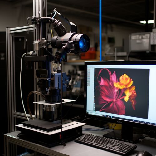
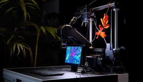
Confocal Laser Scanning Microscopy
Introduction
Confocal Laser Scanning Microscopy (CLSM) is a powerful tool in the field of biological and physical sciences due to its ability to produce in-focus images of thick specimens, a feat unachievable with conventional microscopy techniques. This technique uses a spatial pinhole to eliminate out-of-focus light in specimens that are thicker than the focal plane. This results in sharper images and allows for the collection of serial optical sections from thick specimens.
Principle
The underlying principle of CLSM involves focusing a laser light source onto a single point in a specimen. The light that is reflected or emitted from the specimen is then collected by the objective lens. However, instead of being sent directly to the detector, the light is passed through a spatial pinhole. This pinhole allows only the in-focus light to reach the detector, while the out-of-focus light is blocked. This results in an image that is free from the blurring caused by out-of-focus information.
Components
A typical CLSM system consists of several key components, including a laser light source, scanning mirrors, a spatial pinhole, a detector, and a computer system for image acquisition and processing.
Laser Light Source
The laser light source in a CLSM system is used to illuminate the specimen. The choice of laser and its wavelength is dependent on the specific requirements of the experiment. For example, different lasers may be used for different types of fluorescent dyes.
Scanning Mirrors
Scanning mirrors are used to direct the laser light across the specimen. By rapidly moving the mirrors, the laser light can be scanned across the specimen in a raster pattern, allowing for the collection of an entire image.
Spatial Pinhole
The spatial pinhole is a key component of a CLSM system. It is placed in front of the detector and serves to block out-of-focus light. The size of the pinhole can be adjusted to change the amount of out-of-focus light that is blocked.
Detector
The detector in a CLSM system is used to collect the in-focus light that passes through the pinhole. This light is then converted into an electrical signal that can be processed by the computer system.
Computer System
The computer system in a CLSM system is used for image acquisition and processing. The electrical signals from the detector are converted into digital images, which can then be further processed and analyzed.


Applications
CLSM has a wide range of applications in both the biological and physical sciences. In biology, it is commonly used for imaging thick specimens such as tissue sections or whole organisms. It is also used in the study of cellular structures and processes, such as cell division and migration. In the physical sciences, CLSM is used for the study of materials, including the imaging of surfaces and the measurement of surface roughness.
Advantages and Limitations
CLSM offers several advantages over conventional microscopy techniques. It allows for the imaging of thick specimens and the collection of serial optical sections. It also provides improved image resolution and contrast. However, CLSM also has some limitations. It requires the use of a laser light source, which can be expensive and may cause damage to sensitive specimens. Additionally, the need for a pinhole can limit the amount of light that reaches the detector, reducing the sensitivity of the system.
