Microscopy
Introduction
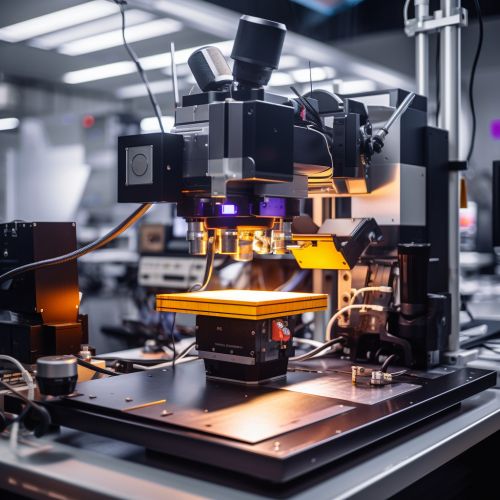
Microscopy is the technical field of using microscopes to view objects and areas of objects that cannot be seen with the naked eye. There are three well-known branches of microscopy: optical, electron, and scanning probe microscopy.
Optical Microscopy
Optical microscopy, also referred to as light microscopy, involves passing visible light transmitted through or reflected from the sample through a single or multiple lenses to allow a magnified view of the sample. The resulting image can be detected directly by the eye, imaged on a photographic plate, or captured digitally.


The most common type of optical microscope is the compound microscope which uses multiple lens elements to increase magnification. The objective lens collects light from the sample, then the ocular lens, or eyepiece lens, magnifies the image. The image can be recorded by means of an attached camera.
Electron Microscopy
Electron microscopy involves the use of an electron microscope that uses a beam of accelerated electrons as a source of illumination. Due to the much smaller wavelength of electrons, electron microscopes have a higher resolving power than light microscopes and can reveal the structure of smaller objects.
A transmission electron microscope (TEM) transmits electrons through a very thinly sliced piece of specimen, while a scanning electron microscope (SEM) scans the sample with a focused electron beam and reads the electrons emitted from the sample. The electron microscope requires vacuum conditions as electrons are easily scattered by molecules in the air.
Scanning Probe Microscopy
Scanning probe microscopy forms images of surfaces using a physical probe that scans the specimen. An image of the surface is obtained by mechanically moving the probe in a raster scan of the specimen, line by line, and recording the probe-surface interaction as a function of position.
The most common types of scanning probe microscopy are atomic force microscopy (AFM) and scanning tunneling microscopy (STM). AFM uses a type of scanning probe microscopy that forms images of surfaces at the atomic level, while STM is a type of microscope used for imaging surfaces at the atomic level.


Applications
Microscopy has been instrumental in the discovery and understanding of the cell, microbiology, and nanotechnology among other fields. It is essential in the field of life sciences, for diagnosing diseases, and in the field of material sciences, for the characterization of materials.
