Two-Photon Excitation Microscopy
Introduction
Two-photon excitation microscopy is a fluorescence imaging technique that allows imaging of living tissue up to about one millimeter in depth. It differs from traditional fluorescence microscopy, in which the excitation wavelength is shorter than the emission wavelength, as the wavelengths of the two exciting photons are longer than the wavelength of the resulting emitted light. This technique is based on the principle of fluorescence, a type of luminescence caused by photons exciting a molecule, raising it to an electronic excited state.


Principle of Operation
In two-photon excitation microscopy, two photons of identical or different frequencies are absorbed, exciting a molecule from one state to a higher energy electronic state. This is a quantum event, the probability of which is quadratic in the excitation intensity, thus it is a low probability event. This process can lead to the emission of a single photon with a wavelength that is shorter than the excitation wavelength, a process known as fluorescence.
The two-photon excitation process can be understood as a simultaneous absorption of two photons by a single molecule. This simultaneous absorption will only occur if the molecule is illuminated with a high density of photons, typically provided by a focused laser beam. The probability of a two-photon absorption event increases with the square of the illumination intensity, thus the rate of two-photon excitation is proportional to the square of the illumination intensity.
Advantages and Applications
Two-photon excitation microscopy has several advantages over traditional fluorescence microscopy. Firstly, the longer excitation wavelength allows for deeper tissue penetration due to reduced scattering. Secondly, the two-photon absorption process is more localized, leading to less photobleaching and photodamage outside the focal volume. Thirdly, the quadratic dependence of the two-photon absorption process on the illumination intensity allows for three-dimensional resolution.
Due to these advantages, two-photon excitation microscopy has found wide applications in the field of biology, especially in imaging living tissues. It is used in neuroscience, developmental biology, and cancer research, among other fields. It allows for the visualization of cells and tissues in their natural state, without the need for slicing or fixing.
Technical Considerations
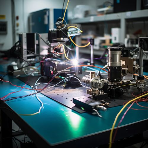
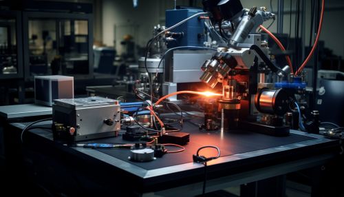
The implementation of two-photon excitation microscopy requires a high-intensity light source, typically a pulsed laser. The laser is focused onto the sample using a high numerical aperture objective. The emitted fluorescence is collected by the same objective and separated from the excitation light by a dichroic mirror. The fluorescence signal is then detected by a photomultiplier tube.
The resolution of two-photon excitation microscopy is determined by the excitation wavelength, the numerical aperture of the objective, and the pulse duration of the laser. The resolution can be improved by using shorter excitation wavelengths, higher numerical aperture objectives, and shorter pulse durations. However, these improvements come at the cost of increased photobleaching and photodamage.
