Diagnostic Imaging
Introduction
Diagnostic imaging, also known as medical imaging, is a discipline within the field of medicine that employs the use of imaging to both diagnose and treat disease visualized within the human body. It encompasses a variety of techniques and procedures that create images of the human body for clinical purposes (medical procedures seeking to reveal, diagnose, or examine disease) or medical science (including the study of normal anatomy and physiology).
History
The history of diagnostic imaging can be traced back to the discovery of radiography by Wilhelm Conrad Roentgen in 1895. This marked the beginning of a new era in medical diagnosis and treatment. Over the years, numerous other imaging modalities have been developed, each with its unique advantages and applications.
Types of Diagnostic Imaging
Diagnostic imaging can be broadly classified into several types, each with its unique set of advantages, limitations, and applications.
Radiography
Radiography involves the use of X-rays to view the internal structures of the body. It is commonly used to diagnose and monitor conditions such as fractures, infections, and tumors. The images produced by radiography are two-dimensional, and the technique is relatively inexpensive and widely available.
Computed Tomography (CT)
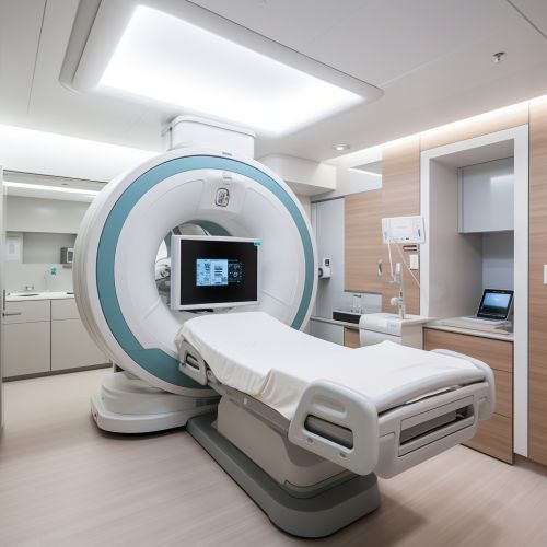
Computed tomography (CT) uses a series of X-ray images taken from different angles around the body and computer processing to create cross-sectional images of the bones, blood vessels, and soft tissues. CT provides greater detail than traditional X-ray exams and can be used to diagnose a variety of conditions, including cancer, heart disease, and trauma injuries.
Magnetic Resonance Imaging (MRI)
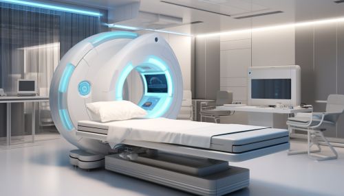
Magnetic resonance imaging (MRI) uses a powerful magnetic field and radio waves to generate detailed images of the organs and tissues within the body. It is particularly useful for imaging soft tissues and organs like the brain, spinal cord, muscles, and heart.
Ultrasound
Ultrasound imaging, also known as sonography, uses high-frequency sound waves to produce images of the inside of the body. It is commonly used during pregnancy to monitor the development of the fetus, but it can also be used to diagnose conditions affecting the heart, blood vessels, and other organs.
Nuclear Medicine
Nuclear medicine involves the use of small amounts of radioactive materials, or radiotracers, to diagnose and treat a variety of diseases. The radiotracers are typically injected into the bloodstream, inhaled, or swallowed, and they emit gamma rays that can be detected by a special camera to create images of the inside of the body.
Applications
Diagnostic imaging plays a crucial role in modern medicine, with applications ranging from diagnosis and treatment planning to research and education.
Diagnosis
Diagnostic imaging is primarily used to diagnose a wide range of conditions, from fractures and infections to cancer and heart disease. The images produced by different imaging modalities can provide detailed information about the structure and function of the body's organs and tissues, helping physicians to identify abnormalities and make accurate diagnoses.
Treatment Planning
In addition to diagnosis, diagnostic imaging is also used to plan and guide treatments. For example, images obtained from CT or MRI can be used to plan radiation therapy for cancer patients, while ultrasound imaging can guide physicians during procedures such as biopsies or catheter placements.
Research and Education
Diagnostic imaging is also an important tool in medical research and education. Researchers use imaging techniques to study the normal anatomy and physiology of the human body, as well as the pathophysiology of disease. In medical education, diagnostic images are used to teach anatomy, demonstrate disease processes, and train future physicians in the interpretation of images.
Safety and Risks
While diagnostic imaging provides invaluable information for the diagnosis and treatment of diseases, it is not without risks. Some imaging modalities, such as radiography and CT, involve exposure to ionizing radiation, which can potentially increase the risk of cancer. However, the benefits of diagnostic imaging generally outweigh the risks, and precautions are taken to minimize radiation exposure.
Future Trends
Advancements in technology and computational power are driving the development of new imaging techniques and the improvement of existing ones. Future trends in diagnostic imaging include the increased use of artificial intelligence to aid in image interpretation, the development of hybrid imaging systems that combine multiple imaging modalities, and the use of molecular imaging to visualize cellular and molecular processes in the body.
See Also


