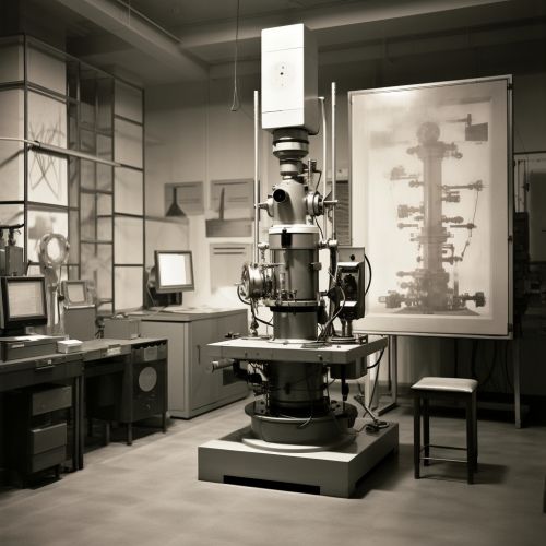
Transmission Electron Microscopy
Introduction
Transmission electron microscopy (TEM) is a microscopy technique that uses a beam of electrons to create an image of the specimen. It is capable of imaging at a significantly higher resolution than light microscopes, allowing it to see fine detail—even as small as a single column of atoms, which are tens of thousands times smaller than a human hair. This makes it an important research tool in fields such as materials science, semiconductor research, and nanotechnology read more.
History
The first transmission electron microscope was developed by Max Knoll and Ernst Ruska in Germany in 1931. Ruska, who was awarded the Nobel Prize in Physics in 1986 for his work, continued to improve the microscope, increasing its resolution from 50 nanometers to less than one nanometer within a few years. This was a significant improvement over optical microscopes, which are limited by the wavelength of visible light.
Principles of Operation
The operation of a TEM is based on the principles of wave-particle duality, which states that particles such as electrons can exhibit properties of both particles and waves. When an electron beam passes through a thin specimen, the electrons are scattered by the atoms in the specimen. This scattering pattern can be captured on a screen to form an image.
Components of a TEM
A typical TEM consists of several key components:
Electron Gun
The electron gun generates the electron beam. It consists of a filament, usually made of tungsten, which is heated to release electrons. These electrons are then accelerated by an electric field to form a beam.
Condenser System
The condenser system focuses the electron beam onto the specimen. It usually consists of two or three electromagnetic lenses.
Specimen Stage
The specimen stage holds the specimen. It can be moved in all three dimensions to allow different areas of the specimen to be examined.
Objective Lens
The objective lens forms the first magnified image of the specimen. It is the most important lens in the microscope, as it determines the resolution and contrast of the image.
Projection System
The projection system further magnifies the image and projects it onto the viewing screen or a camera.
Specimen Preparation
Preparing a specimen for TEM analysis can be a complex process. The specimen must be thin enough to allow electrons to pass through it, typically less than 100 nanometers thick. This often requires the use of specialized techniques such as ultramicrotomy, ion milling, or electro-polishing.
Applications
Due to its high resolution, TEM is used in a wide range of scientific fields. In materials science, it is used to study the structure of materials at the atomic level. In biology, it is used to examine cells and viruses in detail. In semiconductor research, it is used to investigate the structure of semiconductor devices.
Advantages and Limitations
One of the main advantages of TEM is its high resolution, which allows it to see detail at the atomic level. However, it also has several limitations. The preparation of specimens can be time-consuming and complex. The specimens must also be stable under a high vacuum and resistant to damage from the electron beam.
See Also


