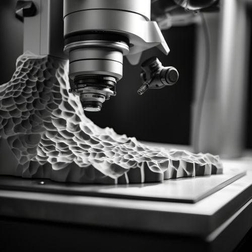
Scanning Electron Microscopy
Introduction
Scanning Electron Microscopy (SEM) is a type of electron microscopy that produces images of a sample by scanning it with a focused beam of electrons. The electrons interact with atoms in the sample, producing various signals that contain information about the sample's surface topography and composition. SEM is widely used in various fields such as materials science, physics, chemistry, biology, geology, and archaeology.
Principle of Operation
The basic principle of SEM involves scanning a sample with a high-energy beam of electrons in a raster scan pattern. The beam's interaction with the sample results in the reflection or emission of electrons and X-rays, which are detected and transformed into an image. The electron beam is produced by an electron gun. This is usually a tungsten filament cathode, although field emission guns (FEGs) are also used. The electron beam is accelerated towards the sample by an anode and passes through electromagnetic lenses, which focus the beam down to a fine point.
Electron Detectors
SEM utilizes various types of detectors to capture different signals produced by the electron-sample interaction. The most common detectors include:
- Secondary Electron Detector (SED): This detector captures secondary electrons, which are ejected from the sample's surface due to the primary electron beam. The SED produces high-resolution, topographic images of the sample.
- Backscattered Electron Detector (BSED): This detector captures backscattered electrons, which are primary beam electrons that have been reflected back from the sample. The BSED produces images that provide information about the sample's composition and crystallographic information.
- Energy-Dispersive X-ray Spectroscopy Detector (EDS): This detector captures X-rays produced by the electron-sample interaction. EDS provides elemental analysis of the sample.
Sample Preparation
Proper sample preparation is crucial for obtaining high-quality SEM images. The preparation method depends on the nature of the sample and the information required. Common preparation techniques include:
- Coating: Non-conductive samples are often coated with a thin layer of conductive material, such as gold, palladium or carbon, to prevent charging under the electron beam.
- Dehydration: Biological samples are usually dehydrated before SEM observation to prevent damage under the electron beam.
- Embedding and Sectioning: Some samples, particularly biological ones, are embedded in resin and sectioned into thin slices for SEM observation.
Applications
SEM has a wide range of applications in various fields:
- In materials science, SEM is used to study the structure, composition, and properties of various materials.
- In biology and medicine, SEM is used to study the detailed morphology of cells and tissues.
- In geology and paleontology, SEM is used to study the morphology and composition of minerals, rocks, and fossils.
- In archaeology, SEM is used to study the composition and manufacturing techniques of ancient artifacts.


See Also
- Transmission Electron Microscopy - Atomic Force Microscopy - X-ray Microanalysis
