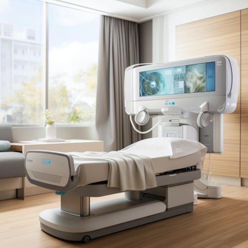
Single Photon Emission Computed Tomography
Introduction
Single Photon Emission Computed Tomography (SPECT) is a type of nuclear medicine imaging technique that provides three-dimensional information about the functional processes within the body. Unlike other imaging techniques such as CT or MRI, which provide anatomical information, SPECT provides functional information about the body's organs and tissues. This makes it a valuable tool in the diagnosis and management of a wide range of medical conditions.


Principles of SPECT
SPECT imaging involves the use of radiopharmaceuticals, which are substances that emit gamma rays when they decay. These radiopharmaceuticals are injected into the patient's bloodstream, where they travel to specific organs or tissues. The gamma rays emitted by the radiopharmaceuticals are detected by a gamma camera, which rotates around the patient to capture images from multiple angles. These images are then reconstructed by a computer to create a three-dimensional image of the organ or tissue being studied.
Applications of SPECT
SPECT imaging has a wide range of applications in clinical medicine. Some of the most common uses of SPECT include:
Cardiology
In cardiology, SPECT is used to assess blood flow to the heart muscle, to detect areas of the heart that are not receiving enough blood, and to assess the function of the heart. This can help in the diagnosis of conditions such as coronary artery disease and myocardial infarction.
Neurology
SPECT is used in neurology to study the brain's function and to detect abnormalities in blood flow or metabolism. This can be useful in the diagnosis of conditions such as stroke, dementia, and epilepsy.
Oncology
In oncology, SPECT can be used to detect and locate tumors, to assess the response to treatment, and to detect recurrence of cancer.
Advantages and Limitations
SPECT imaging has several advantages over other imaging techniques. It provides functional information, which can be more useful than anatomical information in many cases. It can also be used to study a wide range of organs and tissues, and it can provide three-dimensional images.
However, SPECT also has some limitations. The resolution of SPECT images is generally lower than that of CT or MRI images. SPECT also involves exposure to ionizing radiation, which can be a concern for some patients. Finally, the need for radiopharmaceuticals can make SPECT more expensive and less widely available than other imaging techniques.
Future Directions
The field of SPECT imaging is continually evolving, with ongoing research aimed at improving the resolution and sensitivity of SPECT images, reducing the radiation dose, and developing new radiopharmaceuticals. One promising area of research is the development of hybrid imaging techniques, which combine SPECT with other imaging techniques such as CT or MRI to provide both functional and anatomical information.
