Neuroimaging in Rehabilitation
Introduction
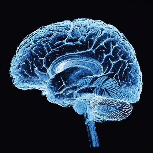
Neuroimaging in rehabilitation is a specialized field of medical science that utilizes various neuroimaging techniques to aid in the assessment and treatment of patients undergoing rehabilitation. This field has seen significant advancements in recent years, with the development of sophisticated imaging technologies that can provide detailed insights into the structure and function of the brain. These technologies have the potential to revolutionize the field of rehabilitation, offering new methods for diagnosing conditions, monitoring patient progress, and developing personalized treatment plans.
Neuroimaging Techniques
There are several neuroimaging techniques commonly used in rehabilitation, each with its own strengths and limitations. These include MRI, fMRI, PET, and DTI, among others.
Magnetic Resonance Imaging (MRI)


MRI is a non-invasive imaging technique that uses a powerful magnetic field and radio waves to generate detailed images of the brain. It is particularly useful in rehabilitation for identifying structural abnormalities in the brain, such as those caused by stroke or traumatic brain injury.
Functional Magnetic Resonance Imaging (fMRI)
fMRI is a variation of MRI that measures brain activity by detecting changes in blood flow. This technique is often used in rehabilitation to assess the functional status of the brain and to monitor changes in brain activity over time.
Positron Emission Tomography (PET)
PET is a nuclear imaging technique that uses radioactive tracers to visualize metabolic processes in the brain. It can provide valuable information about the functional status of the brain, and is often used in rehabilitation to assess the effects of brain injury and to monitor the progress of treatment.
Diffusion Tensor Imaging (DTI)
DTI is a type of MRI that allows for the visualization of white matter tracts in the brain. This technique is particularly useful in rehabilitation for assessing the integrity of these tracts, which can be affected by conditions such as stroke and traumatic brain injury.
Role of Neuroimaging in Rehabilitation
Neuroimaging plays a crucial role in the field of rehabilitation, offering valuable insights into the structure and function of the brain that can inform the development of personalized treatment plans.
Diagnosis
Neuroimaging techniques can be used to diagnose a wide range of conditions that may require rehabilitation, including stroke, traumatic brain injury, and neurodegenerative diseases. By providing detailed images of the brain, these techniques can help clinicians identify the location and extent of brain damage, which can inform the prognosis and guide the treatment plan.
Monitoring Progress
Neuroimaging can also be used to monitor the progress of patients undergoing rehabilitation. By comparing images taken at different points in time, clinicians can assess the effectiveness of treatment and make necessary adjustments to the rehabilitation plan.
Personalized Treatment
Perhaps one of the most exciting applications of neuroimaging in rehabilitation is the potential for personalized treatment. By providing detailed information about the structure and function of an individual's brain, neuroimaging techniques can help clinicians develop treatment plans that are tailored to the specific needs of the patient.
Challenges and Future Directions
While neuroimaging offers many benefits for rehabilitation, there are also several challenges that need to be addressed. These include the high cost of imaging technologies, the need for specialized training to interpret imaging data, and the ethical considerations associated with the use of these technologies.
Despite these challenges, the future of neuroimaging in rehabilitation looks promising. With continued advancements in imaging technologies and the development of new analytical methods, it is likely that the role of neuroimaging in rehabilitation will continue to grow in the coming years.
