Neuroimaging
Introduction
Neuroimaging is a branch of medical imaging that uses various techniques to directly or indirectly image the structure, function, or pharmacology of the nervous system. It is a relatively new discipline within medicine, neuroscience, and psychology.


History
The history of neuroimaging traces back to the early 20th century, with the development of x-ray radiography. However, it wasn't until the introduction of computed tomography (CT) in the 1970s that neuroimaging began to play a significant role in the field of neuroscience.
Techniques
Structural Imaging
Structural imaging, which deals with the structure of the nervous system and the diagnosis of gross intracranial disease, and injury, is used for the diagnosis of large-scale intracranial disease such as tumor and infection. This type of neuroimaging includes CT scans and MRI.

Functional Imaging
Functional imaging, on the other hand, is used to diagnose metabolic diseases and lesions on a finer scale such as Alzheimer's disease, and also for neurological and cognitive psychology research and building brain-computer interfaces. Functional imaging techniques include PET and fMRI.
Molecular Imaging
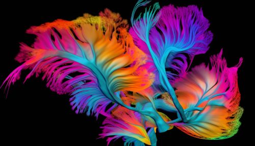
Molecular imaging is a new and expanding discipline which provides detailed visualization of biological processes at the molecular and cellular level in humans and other living systems. Techniques such as MRS and SPECT fall under this category.
Applications
Neuroimaging techniques are used in various fields such as neurology, neuroscience, and psychology. They are used to diagnose diseases and to test the efficacy of treatments. They are also used in research to understand the brain's structure and function, and the relationship between the two.
Future Directions
The future of neuroimaging lies in the development of techniques that can provide more detailed and accurate images of the brain's structure and function. This includes the development of new imaging techniques, as well as the refinement of existing ones.
