Functional Magnetic Resonance Imaging (fMRI)
Introduction
Functional Magnetic Resonance Imaging (fMRI) is a non-invasive neuroimaging technique that allows for the visualization of brain activity. It does this by detecting changes in blood flow in the brain, which are associated with neural activity. This technique has revolutionized the field of neuroscience, providing researchers with an unprecedented view into the human brain at work.
History
The development of fMRI can be traced back to the early 1990s, when it was first introduced as a tool for mapping brain function. It was a significant advancement over previous imaging techniques, such as Positron Emission Tomography (PET), which required the injection of radioactive tracers. The first fMRI studies were conducted using a technique known as Blood Oxygen Level Dependent (BOLD) contrast, which remains the most commonly used method in fMRI research today.
Principles
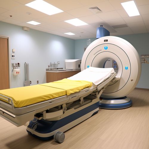
The fundamental principle behind fMRI is the detection of changes in blood flow in the brain. When a particular region of the brain is active, it requires more oxygen to fuel its activity. This increased demand for oxygen is met by an increase in blood flow to the region. This change in blood flow can be detected by the fMRI scanner, which uses a powerful magnetic field to detect changes in the magnetic properties of the blood.
The primary method used in fMRI studies is BOLD contrast. This technique takes advantage of the fact that oxygenated and deoxygenated blood have different magnetic properties. When a region of the brain is active, the increased blood flow to that region leads to an increase in the ratio of oxygenated to deoxygenated blood, which can be detected by the fMRI scanner.


Applications
fMRI has a wide range of applications in both research and clinical settings. In research, it is used to study brain function, including cognitive processes such as attention, memory, and decision-making. It is also used to study the neural basis of various psychiatric and neurological disorders, including schizophrenia, depression, Alzheimer's disease, and Parkinson's disease.
In clinical settings, fMRI is used for pre-surgical planning, to identify areas of the brain that are critical for language, motor function, and other cognitive processes. This helps surgeons avoid damaging these areas during surgery. It is also used to monitor the progress of treatment for various disorders, and to investigate the effects of drugs on brain function.
Limitations
Despite its many advantages, fMRI also has several limitations. One of the main limitations is its relatively low spatial resolution, which means that it can only detect changes in blood flow at a relatively coarse level. This makes it difficult to study the activity of individual neurons or small groups of neurons.
Another limitation is that fMRI measures changes in blood flow, not neural activity directly. This means that it can only provide an indirect measure of brain activity. Furthermore, the relationship between neural activity and changes in blood flow is not fully understood, which can complicate the interpretation of fMRI data.
Finally, fMRI is a relatively expensive technique, which can limit its use in some settings. It also requires a high level of technical expertise to operate the scanner and analyze the data, which can be a barrier to its use.
Future Directions
Despite these limitations, fMRI remains a powerful tool for studying brain function. Future developments in the field are likely to focus on improving the spatial resolution of fMRI, as well as developing new methods for interpreting fMRI data. There is also ongoing research into the use of fMRI for brain-computer interfaces, which could have a wide range of applications in both research and clinical settings.
