Connectome
Overview
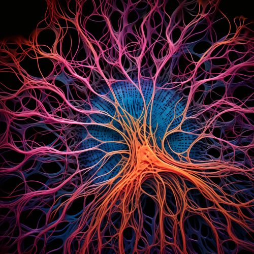
A Connectome is a comprehensive map of neural connections in the brain, and can be thought of as its "wiring diagram". More broadly, a connectome would include the mapping of all neural connections within an organism's nervous system.
History
The term "connectome" was coined by Olaf Sporns and Patric Hagmann in 2005, in the context of their work on structural connectivity in the human brain using diffusion MRI. The term was later popularized by Sebastian Seung in his book "Connectome: How the Brain's Wiring Makes Us Who We Are".


Human Connectome Project
The Human Connectome Project (HCP) is a five-year project sponsored by sixteen components of the National Institutes of Health, split between two consortia of research institutions. The project was launched in July 2009 as the first of a series of projects funded by the NIH Blueprint for Neuroscience Research. The goal of the project is to build a "network map" that will shed light on the anatomical and functional connectivity within the healthy human brain, as well as to produce a body of data that will facilitate research into brain disorders such as dyslexia, autism, Alzheimer's disease, and schizophrenia.
Connectomics
Connectomics is the production and study of connectomes: comprehensive maps of connections within an organism's nervous system. Because these structures are extremely complex, methods have been developed to visualize and analyze them in simpler ways, such as graph theory. Connectomics is the structural study of the number of connections, the connection strength, and the connection length in the brain, and is considered a branch of biomedical imaging and neuroinformatics.
Techniques
There are several techniques currently in use to study the connectome, including:
- Magnetic Resonance Imaging (MRI): This is a non-invasive technique that allows for the visualization of the structure of the brain, and is the most commonly used technique in human connectome studies.
- Diffusion MRI: This is a form of MRI that allows for the imaging of the diffusion process of molecules, mainly water, in biological tissues. In the context of connectomics, diffusion MRI is used to produce images of neural tracts.
- Functional MRI (fMRI): This is another form of MRI that allows for the imaging of brain function. In the context of connectomics, fMRI is used to produce images of brain activity, which can be correlated with the structural connectivity data obtained from diffusion MRI.
- Electroencephalography (EEG) and Magnetoencephalography (MEG): These techniques measure the electrical and magnetic activity of neurons, respectively, and can be used to infer the functional connectivity of the brain.
- Optogenetics: This is a technique that uses light to control neurons that have been genetically modified to express light-sensitive proteins. In the context of connectomics, optogenetics can be used to trace the pathways of specific neural circuits.
Challenges and Criticisms
Despite the potential of connectomics to revolutionize our understanding of the brain, the field has faced several challenges and criticisms. One of the main challenges is the sheer complexity of the brain's connectivity. The human brain is estimated to contain approximately 86 billion neurons, each of which can form thousands of connections with other neurons. This results in an estimated total of 100 trillion connections, all of which would need to be mapped in order to fully understand the brain's connectivity.
Future Directions
The field of connectomics is still in its early stages, and there are many directions in which it could potentially develop. One such direction is the development of new techniques for imaging the brain's connectivity at a higher resolution. Another is the integration of connectomics with other fields of neuroscience, such as genomics and proteomics, in order to gain a more complete understanding of the brain.
