Computerized Tomography
Introduction
Computerized Tomography (CT) is a diagnostic imaging procedure that uses a combination of X-rays and computer technology to produce cross-sectional images (often called slices), both horizontally and vertically, of the body. A CT scan shows detailed images of any part of the body, including the bones, muscles, fat, and organs. CT scans are more detailed than standard X-rays.
History
The development of computerized tomography was made possible by the invention of the computer and advances in digital and detector technology. The first commercial CT scanner was invented by Sir Godfrey Hounsfield in Hayes, United Kingdom, at EMI Central Research Laboratories using X-rays. Hounsfield conceived his idea in 1967, and it was publicly announced in 1972. The first clinical CT scanners were installed between 1974 and 1976. The original systems were dedicated to head imaging only, but "whole body" systems with larger patient openings became available in 1976.


Technology
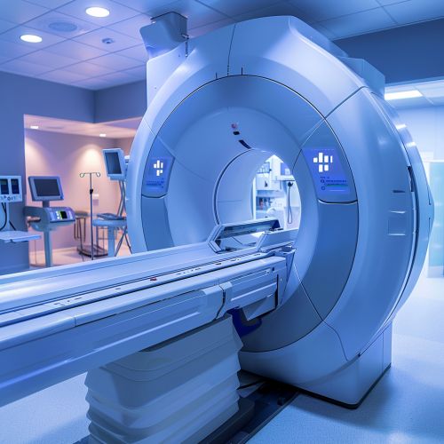
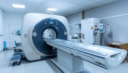
CT scanners are composed of multiple components, including the X-ray generator, X-ray detector, and a system to move the patient through the gantry. The X-ray generator emits a fan-shaped beam of X-rays that pass through the patient and are detected by the X-ray detector. The detector measures the amount of X-rays that make it through the patient and sends this information to the computer.
The computer processes this information to create a two-dimensional cross-sectional image of the patient. The patient is then moved through the gantry and the process is repeated to create another image slice. This process is repeated until the entire area of interest has been imaged.
Procedure
During a CT scan, the patient lies on a table that moves through the gantry of the CT scanner. The X-ray tube rotates around the patient, emitting a fan-shaped beam of X-rays that pass through the patient and are detected by the X-ray detector. The detector sends this information to the computer, which processes it to create a two-dimensional image slice of the patient. The table then moves the patient through the gantry and the process is repeated to create another image slice. This process is repeated until the entire area of interest has been imaged.
Applications
CT scans are used in a variety of medical applications. They can be used to diagnose and monitor a wide range of health conditions, including cancer, heart disease, lung nodules, and liver masses. CT scans can also be used to guide biopsies and other medical procedures, as well as plan for surgeries. In addition, CT scans are used in emergency rooms to diagnose and treat injuries.
Risks and Limitations
While CT scans are a valuable diagnostic tool, they do come with certain risks and limitations. The primary risk associated with CT scans is exposure to ionizing radiation. While the amount of radiation a patient is exposed to during a CT scan is small, repeated exposure can increase the risk of developing cancer.
In addition, there are certain limitations to what a CT scan can detect. For example, CT scans are not as effective at imaging soft tissues as MRI. Furthermore, some patients may be allergic to the contrast materials used in some CT scans.
Future Developments
The field of CT scanning continues to evolve with increasing speed and sophistication. Future developments may include faster scanning times, lower radiation doses, and improved image quality. Additionally, advances in artificial intelligence and machine learning could lead to automated image interpretation, potentially improving diagnostic accuracy and efficiency.
