Brain mapping
Introduction
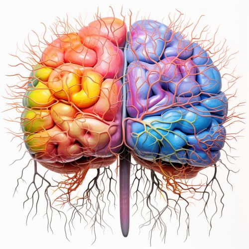
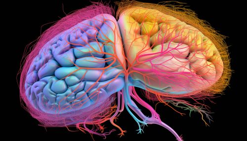
Brain mapping is a set of neuroscience techniques predicated on the mapping of (biological) quantities or properties onto spatial representations of the (human or non-human) brain resulting in maps. This scientific endeavor involves the use of imaging, computational, biophysical, and genetic methods to create spatial representations of the brain's structure, function, and connectivity patterns.
History
The concept of brain mapping has been around for centuries, with early attempts dating back to the ancient Greeks. However, the modern field of brain mapping began to take shape in the late 19th century with the work of pioneers such as Paul Pierre Broca and Carl Ludwig Siegfried Wernicke, who began to link specific brain areas to distinct functions. This early work laid the foundation for the development of more advanced techniques and technologies that have allowed for increasingly detailed maps of the brain's structure and function.


Techniques
Brain mapping techniques are primarily concerned with the study of the anatomy and function of the brain and spinal cord through the use of imaging. This includes various types of imaging techniques such as MRI, fMRI, PET, and Diffusion MRI. These techniques provide detailed images of the brain that are used to create a detailed map of brain structure and connectivity.
Magnetic Resonance Imaging (MRI)
MRI is a non-invasive imaging technology that produces three dimensional detailed anatomical images. It is often used by scientists to image the brain and other parts of the body. It is a useful tool for brain mapping because it provides detailed images of the brain's structure.
Functional Magnetic Resonance Imaging (fMRI)
fMRI is a functional neuroimaging procedure using MRI technology that measures brain activity by detecting changes associated with blood flow. This technique relies on the fact that cerebral blood flow and neuronal activation are coupled. When an area of the brain is in use, blood flow to that region also increases.
Positron Emission Tomography (PET)
PET is a functional imaging technique that uses radioactive substances known as radiotracers to visualize and measure changes in metabolic processes, and in other physiological activities including blood flow, regional chemical composition, and absorption.
Diffusion MRI
Diffusion MRI (or dMRI) is a magnetic resonance imaging method which came into existence in the mid-1980s. It allows the mapping of the diffusion process of molecules, mainly water, in biological tissues, in vivo and non-invasively. Molecular diffusion in tissues is not free, but reflects interactions with many obstacles, such as macromolecules, fibers, membranes, etc.
Applications
Brain mapping techniques are used in various fields of neuroscience, including cognitive neuroscience, cognitive psychology, neuropsychology, and neurobiology. They are also used in clinical settings to diagnose and monitor neurological disorders such as Alzheimer's, Parkinson's, epilepsy, multiple sclerosis, and brain tumors.
Challenges and Future Directions
Despite the advancements in brain mapping techniques, there are still many challenges that need to be addressed. For example, the human brain is incredibly complex, and our understanding of it is still limited. Additionally, there are technical limitations to the current imaging techniques that need to be overcome. However, with the rapid advancements in technology and our growing understanding of the brain, the future of brain mapping looks promising.
