Advances in Brain Mapping Techniques
Introduction
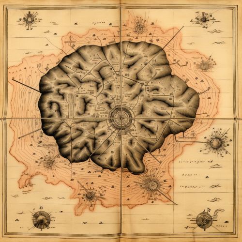
Brain mapping is a set of neuroscience techniques predicated on the mapping of (biological) quantities or properties onto spatial representations of the (human or non-human) brain resulting in maps. All neuroimaging can be considered part of brain mapping. Brain mapping can be conceptualized in various ways: as a literal spatial map of the brain's physical structure, as a metaphorical map of the various features and capacities of the mind, or as a tool for data analysis and visualization. This article will delve into the advances in brain mapping techniques, discussing the evolution, current techniques, and future prospects of this fascinating field of neuroscience.
History of Brain Mapping Techniques
The history of brain mapping techniques is a testament to the relentless pursuit of understanding the human brain. The earliest attempts at brain mapping began with the work of pioneers like Paul Broca and Carl Wernicke, who used lesion studies to localize language functions in the brain. The advent of neuroimaging techniques in the late 20th century, such as computed tomography (CT) and magnetic resonance imaging (MRI), revolutionized brain mapping by allowing for non-invasive visualization of the brain's structure.


Current Techniques
The current landscape of brain mapping techniques is diverse, encompassing structural imaging, functional imaging, and connectomics.
Structural Imaging
Structural imaging techniques provide a static image of the brain's anatomy. The most commonly used structural imaging techniques are MRI and CT scans. MRI uses magnetic fields and radio waves to generate images of the brain, while CT uses X-rays. These techniques have been instrumental in identifying structural abnormalities associated with various neurological disorders.
Functional Imaging
Functional imaging techniques, such as functional MRI (fMRI) and positron emission tomography (PET), measure brain activity by detecting changes associated with blood flow. These techniques have been pivotal in understanding the brain's functional organization, revealing how different brain regions work together to process information and drive behavior.
Connectomics
Connectomics is a research field that aims to map the brain's structural and functional connections. Techniques used in connectomics include diffusion tensor imaging (DTI), which maps white matter tracts, and resting-state fMRI, which maps functional connections between brain regions.
Advances in Brain Mapping Techniques
The field of brain mapping has seen significant advances in recent years, driven by technological innovations and methodological breakthroughs.
High-Resolution Imaging
One of the most significant advances in brain mapping techniques is the development of high-resolution imaging. Techniques such as high-field MRI and ultra-high field MRI (7 Tesla and above) have dramatically increased the spatial resolution of brain images, allowing for more detailed mapping of the brain's structure and function.
Multimodal Imaging
Another major advance is the rise of multimodal imaging, which combines different imaging techniques to provide a more comprehensive view of the brain. For example, fMRI can be combined with DTI to map both the functional and structural connectivity of the brain.
Big Data and Machine Learning
The advent of big data and machine learning has also revolutionized brain mapping. Large-scale neuroimaging datasets, such as the Human Connectome Project, have provided unprecedented opportunities for mapping the human brain. Meanwhile, machine learning algorithms have been increasingly used to analyze these datasets, enabling the discovery of complex patterns and relationships that were previously unattainable.
Future Prospects
The future of brain mapping is promising, with several emerging techniques poised to further advance the field.
Molecular Imaging
Molecular imaging is an emerging field that aims to visualize and quantify molecular processes in the brain. Techniques such as PET and single-photon emission computed tomography (SPECT) can be used to map the distribution of specific molecules in the brain, providing insights into the molecular mechanisms underlying brain function and disease.
Optogenetics
Optogenetics is a technique that uses light to control neurons that have been genetically modified to express light-sensitive proteins. This technique has the potential to provide unprecedented precision in mapping the brain's functional circuits.
Nanotechnology
Nanotechnology holds great promise for advancing brain mapping. For example, nanoparticles can be used as contrast agents in MRI, potentially enhancing the resolution and specificity of brain images.
Conclusion
The field of brain mapping has come a long way since its inception, with advances in technology and methodology continually pushing the boundaries of our understanding of the brain. As we move forward, the integration of different techniques, the application of big data and machine learning, and the exploration of emerging fields like molecular imaging, optogenetics, and nanotechnology will undoubtedly continue to shape the future of brain mapping.
