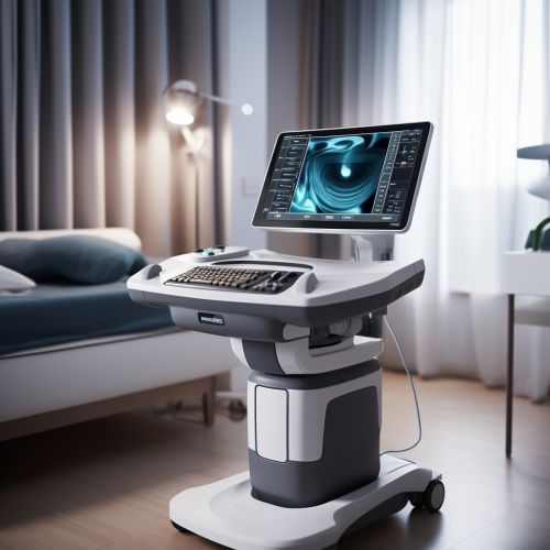
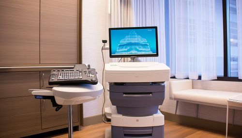
Ultrasound imaging
Introduction
Ultrasound imaging, also known as sonography, is a diagnostic medical procedure that uses high-frequency sound waves to produce dynamic visual images of organs, tissues, or blood flow inside the body. This technique is non-invasive and does not use ionizing radiation, making it a safe tool for medical diagnosis learn more.
History
Ultrasound imaging has a rich history, with its roots in the discovery of piezoelectricity by Pierre and Jacques Curie in 1880. The first practical application of ultrasound technology was in World War I, for the detection of submarines. It was not until the 1940s that ultrasound began to be used in the medical field, with pioneering work by Dr. Karl Theodore Dussik in Austria and Dr. George Ludwig in the United States learn more.


Principles of Ultrasound Imaging
Ultrasound imaging is based on the principles of medical sonography. It involves the transmission of high-frequency sound waves into the body. These sound waves are reflected back to the transducer by the internal structures of the body. The time it takes for the echo to return and the strength of the echo is recorded, creating a two-dimensional image of the internal structure learn more.
Types of Ultrasound Imaging
There are several types of ultrasound imaging, including:
- Doppler Ultrasound: This type of ultrasound is used to measure the speed and direction of blood flow in the body learn more.
- 3D and 4D Ultrasound: These types of ultrasound create three-dimensional images of the fetus in utero. 4D ultrasounds are essentially 3D ultrasounds in motion learn more.
- Echocardiograms: This is a type of ultrasound image of the heart learn more.
- Obstetric Ultrasound: This type of ultrasound is used during pregnancy to monitor the development of the fetus learn more.
Applications of Ultrasound Imaging
Ultrasound imaging has a wide range of applications in the medical field. It is used in obstetrics and gynecology to monitor the development of the fetus and diagnose diseases in the female reproductive system. In cardiology, it is used to diagnose heart conditions and assess damage after a heart attack. Ultrasound imaging is also used in the diagnosis and treatment of conditions in the liver, kidneys, gallbladder, pancreas, thyroid, and other organs learn more.
Advantages and Disadvantages
Like any diagnostic tool, ultrasound imaging has its advantages and disadvantages. One of the main advantages is that it is non-invasive and does not use ionizing radiation, making it safe for pregnant women and patients who cannot undergo certain types of imaging procedures. However, ultrasound images can be difficult to interpret, and the quality of the image depends on the skill of the sonographer learn more.
Future of Ultrasound Imaging
The future of ultrasound imaging is promising, with advancements in technology leading to improved image quality and new applications. Developments in 3D and 4D ultrasound imaging, elastography, and contrast-enhanced ultrasound are paving the way for more accurate diagnoses and treatments learn more.
