Ultrasonography
Introduction
Ultrasonography, also known as ultrasound imaging, is a diagnostic imaging technique that uses high-frequency sound waves to visualize the internal structures of the body. The technique is non-invasive and does not use ionizing radiation, making it a safe tool for medical imaging read more.
History
The application of ultrasound for medical purposes was first explored in the early 20th century. The development of ultrasonography as a diagnostic tool was largely driven by advancements in electronics and computing technology during and after World War II. The first commercially available ultrasound machines were introduced in the 1960s, and the technology has been continually refined and improved since then.
Principles of Ultrasonography
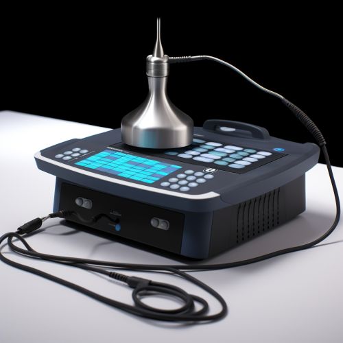
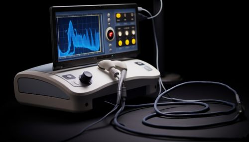
Ultrasonography operates on the principle of the reflection (or echo) of sound waves off internal structures. The ultrasound machine transmits high-frequency sound waves into the body using a transducer. These sound waves travel through the body and bounce back when they encounter different tissues. The returning echoes are picked up by the transducer and are processed by the machine to create an image of the internal structures.
Types of Ultrasonography
There are several types of ultrasonography, each with its specific applications and advantages.
B-Mode Ultrasonography
B-Mode (or brightness mode) ultrasonography is the most common form of ultrasound imaging. It provides a two-dimensional cross-sectional view of the internal structures. B-Mode ultrasonography is widely used in various fields of medicine, including obstetrics, cardiology, and radiology.
Doppler Ultrasonography
Doppler ultrasonography is a specialized form of ultrasound that measures the velocity and direction of blood flow by utilizing the Doppler effect. It is commonly used to detect blood flow problems, such as clots or blockages in the arteries.
3D and 4D Ultrasonography
3D ultrasonography creates three-dimensional images of the internal structures, while 4D ultrasonography adds the element of time to the 3D image, essentially creating a live video of the internal structures. These forms of ultrasonography are particularly useful in obstetrics, where they can provide detailed images of the fetus.
Applications of Ultrasonography
Ultrasonography has a wide range of applications in medical diagnostics. It is commonly used in obstetrics and gynecology to monitor the development of the fetus and to detect any potential abnormalities. In cardiology, ultrasonography is used to assess the structure and function of the heart. It is also used in radiology to visualize the internal organs and to guide interventional procedures.
Advantages and Limitations
Ultrasonography has several advantages over other imaging techniques. It is non-invasive, does not use ionizing radiation, and provides real-time imaging. However, it also has some limitations. The quality of the images can be affected by the patient's body habitus and the presence of gas or bone in the path of the sound waves.


Future Developments
The field of ultrasonography continues to evolve with advancements in technology. Developments in 3D and 4D imaging, elastography, and contrast-enhanced ultrasound are expanding the capabilities of ultrasound imaging. Furthermore, the integration of artificial intelligence and machine learning in ultrasonography is a promising area of research that could potentially improve the accuracy and efficiency of ultrasound imaging.
