Pleural effusion
Introduction
Pleural effusion is a medical condition characterized by the accumulation of excess fluid between the layers of the pleura, the membranes lining the lungs and the chest cavity. This condition can result from various underlying diseases and can significantly impact respiratory function. The pleural space normally contains a small amount of lubricating fluid, but when the balance of fluid production and absorption is disrupted, pleural effusion occurs.
Etiology
Pleural effusion can be classified based on the underlying cause and the nature of the fluid. The two main types are transudative effusion and exudative effusion.
Transudative Effusion
Transudative effusion is usually caused by systemic factors that alter the balance of hydrostatic and oncotic pressures. Common causes include:
- Congestive heart failure: Increased pressure in the pulmonary capillaries leads to fluid leakage into the pleural space.
- Cirrhosis: Reduced albumin production decreases oncotic pressure, promoting fluid accumulation.
- Nephrotic syndrome: Loss of protein in the urine reduces plasma oncotic pressure.
Exudative Effusion
Exudative effusion results from local factors that increase capillary permeability or impair lymphatic drainage. Causes include:
- Infections: Bacterial pneumonia, tuberculosis, and viral infections can cause pleural inflammation and effusion.
- Malignancies: Lung cancer, breast cancer, and lymphoma can invade the pleura or obstruct lymphatic drainage.
- Pulmonary embolism: Infarction and inflammation can lead to pleural effusion.
Pathophysiology
The pathophysiology of pleural effusion involves complex interactions between the pleura, capillaries, and lymphatic system. Normally, the pleural space contains 10-20 mL of fluid, which is maintained by a balance between fluid production and absorption. Disruption of this balance can occur due to:
- Increased capillary hydrostatic pressure
- Decreased plasma oncotic pressure
- Increased capillary permeability
- Impaired lymphatic drainage
The fluid in pleural effusion can be classified as transudate or exudate based on its biochemical characteristics. Light's criteria are commonly used to differentiate between the two types.
Clinical Presentation
Patients with pleural effusion may present with a variety of symptoms, depending on the underlying cause and the volume of fluid. Common symptoms include:
- Dyspnea: Shortness of breath due to lung compression.
- Pleuritic chest pain: Sharp pain exacerbated by breathing or coughing.
- Cough: Non-productive cough due to irritation of the pleura.
Physical examination findings may include:
- Decreased breath sounds
- Dullness to percussion
- Decreased tactile fremitus
Diagnosis
The diagnosis of pleural effusion involves a combination of clinical evaluation, imaging studies, and laboratory analysis.
Imaging Studies
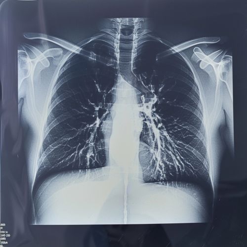
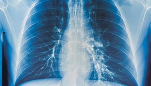
- Chest X-ray: Initial imaging modality to detect pleural effusion. A lateral decubitus view can help assess the volume of fluid.
- Ultrasound: Useful for guiding thoracentesis and assessing fluid characteristics.
- Computed tomography (CT): Provides detailed information about the pleura and underlying lung pathology.
Thoracentesis
Thoracentesis is a procedure in which a needle is inserted into the pleural space to obtain fluid for analysis. The fluid is analyzed for:
- Protein and lactate dehydrogenase (LDH) levels
- Cell count and differential
- pH and glucose levels
- Microbiological cultures
- Cytology for malignant cells
Management
The management of pleural effusion depends on the underlying cause and the severity of symptoms.
Therapeutic Thoracentesis
Therapeutic thoracentesis is performed to relieve symptoms and improve respiratory function. It involves the removal of pleural fluid using a needle or catheter. This procedure can provide immediate relief from dyspnea.
Treatment of Underlying Cause
Addressing the underlying cause is crucial for the long-term management of pleural effusion. This may include:
- Diuretics for congestive heart failure
- Antibiotics for bacterial infections
- Chemotherapy or radiation therapy for malignancies
Pleurodesis
Pleurodesis is a procedure that involves the instillation of a sclerosing agent into the pleural space to induce fibrosis and prevent recurrent effusion. It is commonly used in patients with malignant pleural effusion.
Indwelling Pleural Catheters
Indwelling pleural catheters are used for the management of recurrent pleural effusions, particularly in patients with malignancy. These catheters allow for continuous drainage of pleural fluid at home.
Complications
Complications of pleural effusion can arise from the underlying disease or the treatment itself. Potential complications include:
- Empyema: Infection of the pleural fluid leading to pus accumulation.
- Pneumothorax: Air in the pleural space, often a complication of thoracentesis.
- Fibrothorax: Fibrosis of the pleural space, leading to restricted lung expansion.
Prognosis
The prognosis of pleural effusion depends on the underlying cause and the patient's overall health. Transudative effusions generally have a better prognosis as they are often related to treatable conditions like heart failure. Exudative effusions, particularly those due to malignancy, may have a poorer prognosis.
See Also
References


