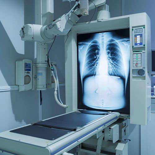
Chest X-ray
Introduction
A Chest X-ray is a common imaging test that uses small amounts of radiation to produce pictures of the organs, tissues, and bones of the chest. This test is often used to diagnose and monitor conditions such as pneumonia, lung cancer, heart conditions, and chest injuries.
Procedure
The chest X-ray procedure is a quick and painless test that typically takes only a few minutes to complete. The patient is asked to stand or sit in front of a special machine that emits X-rays, a type of radiation that can pass through most objects, including the body. The X-rays are absorbed in different amounts by different parts of the body. The X-rays that pass through the body are captured on a detector or film on the other side of the patient, creating an image of the inside of the chest.
Interpretation
Interpreting a chest X-ray requires a thorough understanding of anatomy, physiology, and pathology. The radiologist, a doctor who specializes in interpreting medical images, will look for abnormalities in the lungs, heart, bones, and other structures in the chest. Abnormal findings may include signs of infection, tumors, fractures, fluid in the lungs, and changes in the size or shape of the heart.
Uses
Chest X-rays are used for a variety of purposes. They can help diagnose lung conditions such as pneumonia, tuberculosis, and lung cancer. They can also be used to evaluate the heart, including the size and shape of the heart and the condition of the blood vessels. Chest X-rays can also reveal problems with the bones and soft tissues in the chest, such as fractures and tumors.
Risks
Like all medical procedures, chest X-rays carry some risks. The amount of radiation used in a chest X-ray is small, but repeated exposure can increase the risk of developing cancer. Pregnant women are advised to avoid X-rays because the radiation can harm the developing fetus. Other risks include allergic reactions to contrast materials used in some types of X-rays.
Limitations
While chest X-rays are a valuable diagnostic tool, they have some limitations. They provide a two-dimensional image of a three-dimensional object, which can make it difficult to detect certain conditions. They also cannot provide detailed images of soft tissues, such as the lungs and heart, which can limit their usefulness in diagnosing certain conditions.
Future Developments
Advancements in imaging technology are continually improving the quality and usefulness of chest X-rays. Digital radiography, for example, allows for immediate image review and eliminates the need for film. Other advancements include the use of computer algorithms to help interpret images and the development of portable X-ray machines that can be used in remote locations.


