Neuroanatomy
Introduction
Neuroanatomy is the study of the structure and organization of the nervous system. Compared to other organs, the nervous system is highly complex and involves a multitude of specialized structures and unique biological processes. This article delves into the intricate details of neuroanatomy, shedding light on the various aspects of the nervous system, from the cellular level to the system level.
Structure of the Nervous System
The nervous system can be divided into two main parts: the central nervous system (CNS) and the peripheral nervous system (PNS). The CNS consists of the brain and spinal cord, while the PNS includes all the nerves that connect the CNS to the rest of the body.


Central Nervous System
The central nervous system is the command center of the body, receiving information from the peripheral nervous system, processing it, and sending out instructions. It is composed of the brain and spinal cord.
Brain
The brain is the most complex organ in the human body. It is divided into several regions, each with specific functions and structures. These include the cerebrum, cerebellum, and brainstem.
Cerebrum
The cerebrum is the largest part of the brain. It is divided into two hemispheres and is responsible for higher brain functions such as thought, emotion, and voluntary movement. Each hemisphere is further divided into four lobes: the frontal lobe, parietal lobe, occipital lobe, and temporal lobe.
Cerebellum
The cerebellum is located at the back of the brain, below the cerebrum. It plays a crucial role in motor control, coordination, precision, and accurate timing.
Brainstem
The brainstem connects the cerebrum and cerebellum to the spinal cord. It controls many basic functions, including heart rate, breathing, and sleep.
Spinal Cord
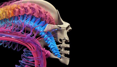
The spinal cord is a long, thin, tubular structure that extends from the brainstem to the lower back. It carries signals between the brain and the rest of the body.
Peripheral Nervous System
The peripheral nervous system consists of all the nerves outside the central nervous system. It is divided into the somatic nervous system, which controls voluntary muscle movement, and the autonomic nervous system, which controls involuntary functions.
Neurons and Glial Cells
The nervous system is made up of two main types of cells: neurons and glial cells. Neurons are the primary cells of the nervous system, responsible for transmitting information throughout the body. Glial cells, on the other hand, provide support and protection for neurons.
Neuroanatomical Techniques
Several techniques are used in neuroanatomy to study the structure of the nervous system. These include histology, magnetic resonance imaging (MRI), and functional MRI (fMRI).
Conclusion
Understanding neuroanatomy is essential for a comprehensive understanding of how the nervous system functions. It provides a foundation for exploring the complex interactions that occur within the nervous system, leading to our thoughts, emotions, and behaviors.
