Musculoskeletal ultrasound
Introduction
Musculoskeletal ultrasound (MSK US) is a non-invasive diagnostic technique used to visualize muscles, tendons, ligaments, nerves, and joints throughout the body. This imaging modality uses ultrasound waves to produce images of the musculoskeletal system, providing valuable information about the anatomy and pathology of these structures.


Principles of Musculoskeletal Ultrasound
MSK US operates on the principles of medical ultrasound. It utilizes high-frequency sound waves, beyond the range of human hearing, to create images of the body's internal structures. The ultrasound machine sends these sound waves into the body using a transducer, a handheld device that is placed on the skin over the area of interest. The sound waves then bounce back, or echo, off the tissues and structures within the body. These echoes are picked up by the transducer and sent back to the machine, which translates them into an image.
Indications for Musculoskeletal Ultrasound
MSK US can be used to diagnose a wide range of conditions affecting the musculoskeletal system. These include, but are not limited to:
- Tendinopathies such as tendonitis and tendon tears
- Muscle strains and tears
- Ligament sprains and tears
- Bursitis
- Arthritic conditions
- Ganglion cysts
- Hernias
- Nerve entrapments and neuropathies
- Soft tissue tumors
Advantages and Limitations
MSK US has several advantages over other imaging modalities. It is non-invasive and does not use radiation, making it safe for all patients, including pregnant women and children. It provides real-time imaging, allowing for dynamic assessment of the musculoskeletal system. It can also provide a multiplanar view of the structures, which is not possible with plain radiographs.
However, MSK US also has its limitations. It is operator-dependent, meaning the quality of the images and the accuracy of the diagnosis can vary based on the skill and experience of the sonographer. It may not be as effective in imaging deep structures or those obscured by bone. Furthermore, it may not provide as detailed an image as MRI, particularly for small structures and intra-articular pathology.
Technique
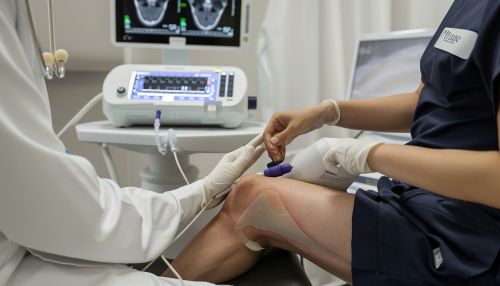
The technique for performing MSK US depends on the specific structure or area of the body being examined. However, there are some general steps that are typically followed. The patient is positioned so that the area of interest is accessible and comfortable. The skin over the area is cleaned, and a water-based gel is applied to help the transducer make good contact with the skin and improve the quality of the images. The sonographer then moves the transducer over the area, adjusting the angle and pressure as needed to obtain the best images.
Interpretation of Results
Interpreting the results of MSK US requires a thorough understanding of the anatomy and pathology of the musculoskeletal system. The sonographer will look for any abnormalities in the size, shape, and echo pattern of the structures. These may indicate conditions such as inflammation, tears, tumors, or other abnormalities.
Future Directions
Advancements in ultrasound technology and techniques continue to improve the utility of MSK US. Three-dimensional ultrasound, elastography, and contrast-enhanced ultrasound are some of the emerging techniques that may provide more detailed and accurate imaging of the musculoskeletal system.
