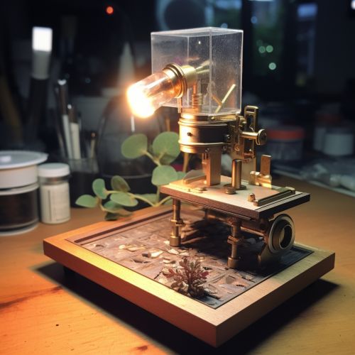
Microscope
Introduction
A microscope is a scientific instrument used to magnify objects that are too small to be seen by the naked eye. The term microscope is derived from the Greek words "micros" meaning small and "skopein" meaning to look or see. Microscopes can be broadly classified into light microscopes and electron microscopes, each with its own set of advantages and disadvantages.


History
The microscope was invented in the 17th century by Dutch spectacle makers, Hans Lippershey, Zacharias Jansen, and his father Hans Jansen. The first microscope consisted of a tube with a plate for the object at one end and a viewing point at the other. This basic design, known as a compound microscope, has evolved over time into the sophisticated instruments we use today.
Types of Microscopes
Microscopes can be categorized based on the type of technology they use to create an image. The two main types are light microscopes and electron microscopes.
Light Microscopes
Light microscopes, also known as optical microscopes, use visible light and a system of lenses to magnify images of small samples. There are several types of light microscopes, including compound microscopes, stereo microscopes, and confocal microscopes.
Compound Microscopes
A compound microscope uses a series of lenses to magnify an object. The object is illuminated from below and viewed through an eyepiece at the top of the microscope. Compound microscopes can achieve magnifications of up to 2000x.
Stereo Microscopes
A stereo microscope, also known as a dissecting microscope, provides a three-dimensional view of the sample. It uses two separate optical paths with two eyepieces to create a depth perception. Stereo microscopes are often used for dissection, microsurgery, watch-making, small circuit board manufacture or inspection, and the like.
Confocal Microscopes
A confocal microscope uses a laser to scan a specimen, which is then reconstructed with a computer to provide a three-dimensional image. Confocal microscopes are used in biological and medical research.
Electron Microscopes
Electron microscopes use a beam of electrons instead of light to magnify the sample. They provide much higher magnification and resolution than light microscopes, allowing scientists to view structures at the molecular level. There are two main types of electron microscopes: transmission electron microscopes (TEMs) and scanning electron microscopes (SEMs).
Transmission Electron Microscopes (TEMs)
TEMs operate on the same basic principles as light microscopes but use electrons instead of light waves. They can magnify images up to a million times their original size.
Scanning Electron Microscopes (SEMs)
SEMs create images by bouncing electrons off the surface of a sample. This produces a detailed, three-dimensional image of the sample's surface topography.
Applications
Microscopes have a wide range of applications in various fields, including biology, medicine, materials science, chemistry, and geology.
Biology and Medicine
In biology and medicine, microscopes are used to study cells and tissues, to diagnose diseases, and to conduct research. For example, pathologists use microscopes to examine tissue samples and identify abnormalities.
Materials Science
In materials science, microscopes are used to study the structure and properties of materials at the microscopic level. This can help scientists understand how materials behave under different conditions and how they can be improved.
Chemistry
In chemistry, microscopes are used to study the structure and behavior of atoms and molecules. This can help scientists understand chemical reactions and the properties of different substances.
Geology
In geology, microscopes are used to study rocks, minerals, and other earth materials. This can help scientists understand the history of the Earth and the processes that shape its surface.
Future Developments
The field of microscopy continues to evolve, with new technologies and techniques being developed all the time. For example, super-resolution microscopy is a recent development that allows scientists to view structures smaller than the diffraction limit of light. Another promising area of research is in the field of quantum microscopy, which uses the principles of quantum mechanics to achieve even higher levels of detail and precision.
