Mandibular Canal
Anatomy
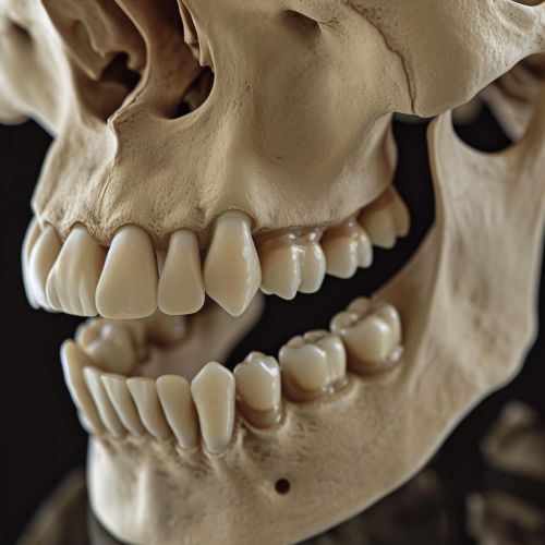
The mandibular canal is a vital anatomical structure in the lower jaw, or the mandible. It is a narrow, tube-like passage that runs bilaterally within the mandible, starting from the mandibular foramen and ending at the mental foramen. The canal houses the inferior alveolar nerve, artery, and vein, which are essential for the sensory and blood supply to the lower teeth and gingiva.


Structure
The mandibular canal begins at the mandibular foramen, located on the inner surface of the ramus of the mandible. It runs obliquely downward and forward in the ramus, and then horizontally forward in the body of the mandible, below the apices of the teeth. The canal finally terminates at the mental foramen, which opens on the outer surface of the body of the mandible.
Contents
The mandibular canal contains the inferior alveolar nerve, artery, and vein. The inferior alveolar nerve is a branch of the trigeminal nerve and provides sensory innervation to the lower teeth, lower lip, and chin. The inferior alveolar artery and vein are branches of the maxillary artery and vein, respectively, and provide blood supply to the lower teeth and gingiva.
Clinical Significance
The mandibular canal is of significant clinical importance in dentistry and oral surgery. Knowledge of its anatomy is crucial during procedures such as the extraction of lower teeth, placement of dental implants, and administration of local anesthesia in the lower jaw. Damage to the canal or its contents during these procedures can lead to complications such as bleeding, infection, or paresthesia (numbness) of the lower lip and chin.
Variations
Anatomical variations of the mandibular canal are not uncommon and can pose challenges during dental procedures. These variations can include bifurcation (splitting into two) of the canal, presence of accessory (additional) canals, and variations in the location and size of the mental foramen. Preoperative imaging, such as dental cone beam computed tomography, is often used to identify these variations and plan the surgical procedure accordingly.
