Mandible
Anatomy
The mandible, also known as the lower jaw, is a bone located in the facial structure of vertebrates, including humans. It is the largest and strongest bone in the human face, playing a vital role in facial aesthetics and function. The mandible is responsible for the movement of the jaw, enabling actions such as chewing, speaking, and facial expressions.


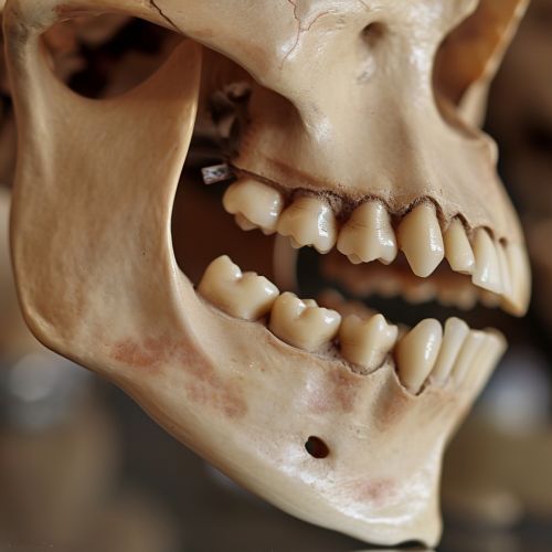
The mandible is a U-shaped bone, with two major parts: the body and the ramus. The body is the horizontal portion that forms the chin and holds the lower teeth, while the ramus is the vertical portion that connects the body of the mandible to the temporal bone of the skull, forming the temporomandibular joint (TMJ).
Body of the Mandible
The body of the mandible is a curved, horizontal structure that forms the base of the lower jaw. It consists of an outer and inner surface, as well as superior and inferior borders. The outer surface of the body is slightly convex and contains several important landmarks, including the mental protuberance (chin) and the mental foramen, which allows passage of the mental nerve and vessels.
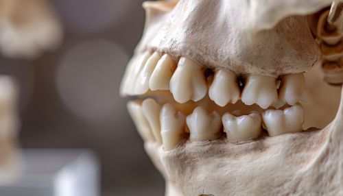
The inner surface of the body contains the mylohyoid line, which is a ridge of bone that serves as the attachment site for the mylohyoid muscle, a muscle that plays a crucial role in swallowing and speech. The superior border of the body contains sockets, known as alveoli, for the roots of the lower teeth. The inferior border is thicker and provides attachment for muscles such as the platysma and depressor labii inferioris.
Ramus of the Mandible
The ramus of the mandible is a quadrilateral-shaped structure that extends upward from the body of the mandible. It has two surfaces (medial and lateral) and four borders (superior, inferior, anterior, and posterior). The superior border of the ramus is the most complex, as it splits into two processes: the coronoid process and the condylar process.
The coronoid process is a thin, triangular eminence that serves as the insertion point for the temporalis muscle, a muscle involved in jaw closure. The condylar process, on the other hand, ends in the mandibular condyle, which articulates with the temporal bone to form the TMJ. This joint allows for the movement of the mandible during mastication and speech.
Development
The development of the mandible begins during the embryonic period, with the formation of the first pharyngeal arch, also known as the mandibular arch. This arch gives rise to two cartilaginous structures, the Meckel's cartilages, which serve as the initial framework for the mandible.
By the end of the embryonic period, intramembranous ossification - a process where bone tissue develops directly from mesenchyme - begins to occur around the Meckel's cartilages. This process forms the body and ramus of the mandible. The Meckel's cartilages eventually degenerate, except for a small portion that contributes to the formation of the malleus and incus, two bones in the middle ear.
Clinical Significance
Due to its prominent location and function, the mandible is often involved in various clinical conditions, ranging from congenital anomalies to traumatic injuries and diseases.
One of the most common congenital anomalies of the mandible is micrognathia, a condition where the mandible is undersized. This condition can lead to difficulties in feeding, breathing, and speech. Treatment options for micrognathia include orthodontic treatment, surgical intervention, or a combination of both.
Traumatic injuries to the mandible, such as fractures, are often caused by physical assault, motor vehicle accidents, or sports injuries. These injuries can lead to severe pain, difficulty in opening the mouth, and facial deformity. Treatment often involves surgical intervention to realign and stabilize the fractured segments.
Diseases that can affect the mandible include osteomyelitis (infection of the bone), osteonecrosis (death of bone tissue), and tumors, both benign and malignant. These conditions often require medical or surgical treatment, depending on the severity and extent of the disease.
