Magnetic Resonance Imaging Scans
Introduction
Magnetic Resonance Imaging (MRI) is a non-invasive imaging technology that produces three dimensional detailed anatomical images. It is often used for disease detection, diagnosis, and treatment monitoring. It is based on sophisticated technology that excites and detects the change in direction of the rotational axis of protons found in the water that makes up living tissues Learn more about Protons.
Physics of MRI
MRI scanners use strong magnetic fields, magnetic field gradients, and radio waves to generate images of the organs in the body. The MRI scanner can detect signals from different types of atoms, but it is most sensitive to hydrogen, the most abundant atom in the body. The strong magnetic field aligns the nuclear magnetization of (usually) hydrogen atoms in water in the body. Radio frequency fields are used to systematically alter the alignment of this magnetization, causing the hydrogen nuclei to produce a rotating magnetic field detectable by the scanner. This signal can be manipulated by additional magnetic fields to build up enough information to construct an image of the body.
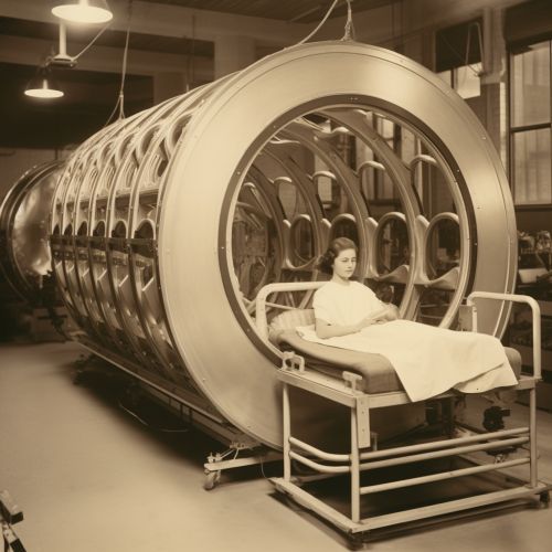
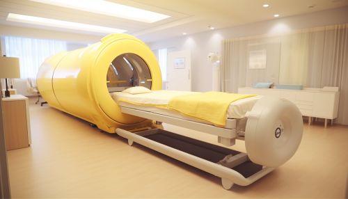
Components of an MRI System
An MRI system is made up of several key components, including the main magnet, gradient system, RF system, patient table, and computer system.
Main Magnet
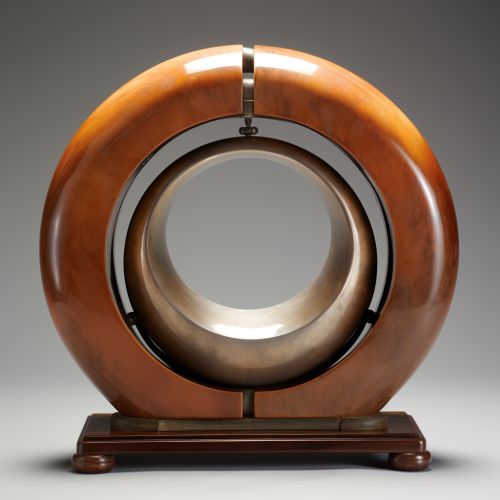
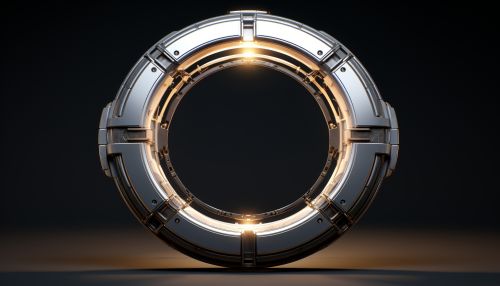
The main magnet is the largest and most obvious component of the MRI system. It creates a strong, stable magnetic field around the patient. The strength of the magnetic field is measured in Tesla (T). Most clinical MRI systems are 1.5T or 3T, but higher field strengths are available for research applications.
Gradient System
The gradient system consists of three smaller coils within the main magnet. These create a secondary magnetic field that varies in strength in a precise, linear way. This allows the scanner to select a specific 'slice' of the patient to image.
RF System
The RF system includes the RF transmitter, which sends the radio frequency pulses to the patient, and the RF receiver, which detects the signal returned from the patient. The RF coils can be built into the scanner or they can be separate devices that are placed around the part of the body being imaged.
Patient Table
The patient table is a motorized bed that moves the patient into and out of the scanner. It can be adjusted to position the patient correctly for the scan.
Computer System
The computer system controls the other components of the MRI system, processes the data, and reconstructs it into images. It also includes the user interface, which allows the technologist to control the scan parameters.
MRI Procedure
During an MRI scan, the patient lies on the patient table which then moves into the scanner. The technologist operates the scanner from a separate room where they can see and hear the patient. The scan can take anywhere from 15 to 90 minutes, depending on the type of scan. The patient must remain still during the scan to avoid blurring the images.


Applications of MRI
MRI has a wide range of applications in medical diagnosis and can be used to image any part of the body. Some of the most common uses include imaging of the brain, spine, joints, abdomen, blood vessels, and heart.
Brain Imaging
MRI is the most commonly used imaging modality for the brain. It can detect a variety of conditions of the brain including tumors, stroke, dementia, and multiple sclerosis.
Spine Imaging
MRI is also commonly used for imaging the spine. It can detect conditions such as spinal cord injury, disc herniation, spinal tumors, and spinal infection.
Joint Imaging
MRI is often used for imaging the joints, including the knee, shoulder, hip, elbow, and wrist. It can detect a range of conditions including arthritis, cartilage damage, ligament tears, and bone fractures.
Abdominal Imaging
MRI can be used to image the organs of the abdomen, including the liver, kidneys, pancreas, and adrenal glands. It can detect tumors, cysts, and other abnormalities.
Vascular Imaging
MRI can also be used to image the blood vessels, in a procedure called Magnetic Resonance Angiography (MRA). This can detect problems such as aneurysms, blockages, and malformations.
Cardiac Imaging
Cardiac MRI can image the heart and the surrounding blood vessels. It can detect conditions such as coronary artery disease, heart failure, and congenital heart defects.
Safety and Contraindications
MRI is generally a safe procedure. However, because it uses a strong magnetic field, it is not suitable for people with certain types of metal implants, such as pacemakers, cochlear implants, certain types of vascular clips, or metal fragments in their eyes. Pregnant women should also avoid MRI during their first trimester unless absolutely necessary. Some people may feel claustrophobic in the scanner and may require sedation.
Future of MRI
MRI technology continues to evolve, with ongoing research into new applications and techniques. Some areas of research include functional MRI (fMRI), which can measure brain activity, and diffusion tensor imaging (DTI), which can map the white matter tracts of the brain. Other areas of research include the development of new contrast agents, faster scanning techniques, and the use of AI in image interpretation.


See Also
Functional MRI (fMRI) Diffusion Tensor Imaging (DTI) AI in Radiology
