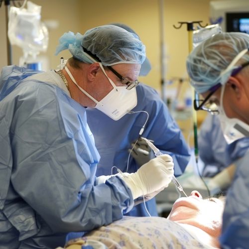
Lung biopsy
Introduction
A lung biopsy is a medical procedure in which a small sample of lung tissue is removed and examined under a microscope to diagnose lung diseases. This procedure is often used to diagnose conditions such as lung cancer, infections, and interstitial lung disease. The biopsy can be performed using various techniques, each with its own indications, advantages, and risks.
Indications
Lung biopsies are indicated in several clinical scenarios. These include:
- **Diagnosis of Lung Cancer**: When imaging studies such as CT scans or PET scans reveal a suspicious lesion, a biopsy is often required to confirm malignancy and determine the type of lung cancer.
- **Evaluation of Interstitial Lung Disease**: Conditions like idiopathic pulmonary fibrosis and sarcoidosis often require a biopsy for definitive diagnosis.
- **Investigation of Infections**: In cases where infections such as tuberculosis or fungal infections are suspected, especially in immunocompromised patients, a biopsy can provide a definitive diagnosis.
- **Assessment of Lung Nodules**: Small nodules detected on imaging studies may require biopsy to determine if they are benign or malignant.
Techniques
There are several techniques for performing a lung biopsy, each with specific indications and contraindications.
Bronchoscopic Biopsy
A bronchoscopic biopsy involves the use of a bronchoscope, a flexible tube with a camera and tools, to obtain tissue samples from the lungs. This technique is less invasive and is often used for lesions near the airways.
- **Indications**: Central lung lesions, suspected infections, and diffuse lung diseases.
- **Advantages**: Minimally invasive, can be performed under local anesthesia.
- **Risks**: Bleeding, infection, pneumothorax.
Needle Biopsy
A needle biopsy, also known as a percutaneous biopsy, involves the insertion of a needle through the chest wall to obtain a tissue sample. This procedure is often guided by imaging techniques such as CT or ultrasound.
- **Indications**: Peripheral lung lesions, solitary pulmonary nodules.
- **Advantages**: Less invasive than surgical biopsy, high diagnostic yield.
- **Risks**: Pneumothorax, bleeding, infection.
Surgical Biopsy
Surgical biopsies are more invasive and are typically performed when other methods are inconclusive or not feasible. There are two main types: open lung biopsy and video-assisted thoracoscopic surgery (VATS).
- **Open Lung Biopsy**: Involves a thoracotomy, where a surgeon makes an incision in the chest to access the lung tissue.
- **VATS**: A minimally invasive technique that uses small incisions and a thoracoscope to obtain tissue samples.
- **Indications**: Diffuse lung diseases, inconclusive results from less invasive methods.
- **Advantages**: High diagnostic accuracy.
- **Risks**: Higher morbidity, longer recovery time.
Procedure
The specific steps of a lung biopsy depend on the technique used. However, general steps include:
1. **Pre-procedure Evaluation**: Includes a thorough medical history, physical examination, and imaging studies to locate the lesion. 2. **Anesthesia**: Local anesthesia for bronchoscopic and needle biopsies; general anesthesia for surgical biopsies. 3. **Tissue Sampling**: Using the chosen technique, tissue samples are obtained and sent to a pathology lab for analysis. 4. **Post-procedure Care**: Monitoring for complications such as bleeding or pneumothorax, and managing pain.
Complications
While lung biopsies are generally safe, they do carry risks. Common complications include:
- **Pneumothorax**: The most common complication, especially with needle biopsies. It involves air leaking into the space between the lung and chest wall, causing the lung to collapse.
- **Bleeding**: Can occur at the biopsy site and may require intervention if severe.
- **Infection**: Although rare, infections can occur and may require antibiotic treatment.
- **Pain**: Post-procedure pain is common and can usually be managed with analgesics.
Pathological Examination
The tissue obtained from a lung biopsy is examined by a pathologist. The examination includes:
- **Histopathology**: Microscopic examination of tissue architecture and cellular details.
- **Immunohistochemistry**: Uses antibodies to detect specific antigens in the tissue, aiding in the diagnosis of certain types of lung cancer.
- **Molecular Testing**: Techniques such as PCR and next-generation sequencing can identify genetic mutations and infections.
Clinical Implications
The results of a lung biopsy can have significant clinical implications:
- **Cancer Diagnosis**: Determines the type and stage of lung cancer, guiding treatment decisions.
- **Infection Identification**: Confirms the presence of infectious organisms, guiding antimicrobial therapy.
- **Interstitial Lung Disease**: Provides a definitive diagnosis, guiding management and prognosis.
Conclusion
Lung biopsy is a critical diagnostic tool in the evaluation of various lung diseases. The choice of biopsy technique depends on the clinical scenario, location of the lesion, and patient's overall health. While the procedure carries risks, its benefits in providing a definitive diagnosis often outweigh the potential complications.
See Also
- Bronchoscopy
- Computed Tomography
- Idiopathic Pulmonary Fibrosis
- Positron Emission Tomography
- Sarcoidosis
- Ultrasound


