Iris (anatomy)
Anatomy
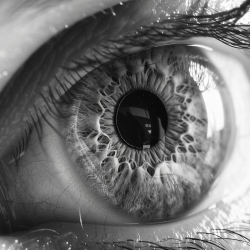
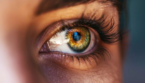
The human eye is a complex organ, and one of its most important components is the iris. The iris is a thin, circular structure in the eye, responsible for controlling the diameter and size of the pupil, and thus the amount of light reaching the retina. The iris is located in front of the lens and the ciliary body, and behind the cornea.


Structure
The iris is composed of two layers: the front pigmented fibrovascular known as a stroma and, beneath the stroma, pigmented epithelial cells. The stroma connects to a sphincter muscle (sphincter pupillae), which contracts the pupil in a circular motion, and a set of dilator muscles (dilator pupillae) which pull the iris radially to enlarge the pupil, pulling it in folds.
Function
The primary function of the iris is to control the amount of light that enters the eye. It does this by adjusting the size of the pupil, the hole at the centre of the iris through which light enters the eye. This is known as the pupillary light reflex.
Clinical significance
Various diseases and conditions can affect the iris, leading to changes in eye color, shape, or function. These include Iritis, Aniridia, and Coloboma. Iritis is inflammation of the iris, while aniridia is a rare congenital condition characterized by the absence of the iris. Coloboma is a hole in one of the structures of the eye, such as the iris, that is present from birth.
