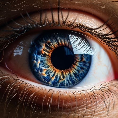
Human eye
Anatomy of the Human Eye
The human eye is a complex organ that provides us with the sense of sight, enabling us to perceive and interpret the shapes, colors, and dimensions of objects in the world by processing the light they reflect or emit. The eye is part of the sensory nervous system.
The eye is not shaped like a perfect sphere, rather it is a fused two-piece unit. The smaller frontal unit, more curved, called the cornea is linked to the larger unit called the sclera. The cornea allows light to enter the eye. As light passes through the eye, the iris changes shape by expanding and letting less light through or contracting and letting more light through to change pupil size. The lens then adjusts to the light passed through the pupil to properly focus it on the retina. The retina then goes on to send this focused image to the brain for processing.


Cornea
The cornea is the transparent front part of the eye that covers the iris, pupil, and anterior chamber. The corneal tissue is arranged in five basic layers, each having an important function. These five layers are: the corneal epithelium, Bowman's layer, the corneal stroma, Descemet's membrane, and the corneal endothelium.
Iris
The iris is a thin, circular structure in the eye, responsible for controlling the diameter and size of the pupil and thus the amount of light reaching the retina. The color of the iris gives the eye its color.
Pupil
The pupil is a hole located in the center of the iris of the eye that allows light to strike the retina. Its function is to control the amount of light entering the eye by changing its size; it constricts (gets smaller) in response to bright light and dilates (gets larger) in response to dim light.
Lens
The lens is a transparent, biconvex structure in the eye that, along with the cornea, helps to refract light to be focused on the retina. By changing shape, it changes the focal distance of the eye so that it can focus on objects at various distances, allowing a sharp real image of the object of interest to be formed on the retina.
Retina
The retina is a thin layer of tissue that lines the back of the eye on the inside. It is composed of several layers, including one that contains specialized cells called photoreceptors. There are two types of photoreceptor cells in the human retina—rods and cones. Rods function mainly in dim light and provide black-and-white vision, while cones support daytime vision and the perception of color.
Function of the Human Eye
The primary function of the eye is to receive light and convert it into electrical signals that the brain can interpret as visual images. This process, known as visual transduction, occurs in the retina and involves several stages.
Light Reception
The first stage of visual transduction is light reception, which occurs when photons enter the eye and strike the photoreceptor cells of the retina. The cornea and lens focus the light onto the retina, where it is absorbed by the photopigments in the photoreceptor cells.
Signal Conversion and Transmission
The absorption of light by the photopigments triggers a chemical reaction that results in the generation of an electrical signal. This signal is then transmitted from the photoreceptor cells to the bipolar cells in the retina, and from there to the ganglion cells.
Signal Processing
The ganglion cells process the electrical signals from the photoreceptors and bipolar cells and generate action potentials, which are transmitted along the optic nerve to the brain. The brain then interprets these signals as visual images.
Diseases and Disorders of the Human Eye
There are many diseases and disorders that can affect the human eye. Some of these include cataracts, glaucoma, macular degeneration, and diabetic retinopathy.
Cataracts
A cataract is a clouding of the lens in the eye that affects vision. Most cataracts are related to aging and are very common in older people.
Glaucoma
Glaucoma is a group of diseases that damage the eye's optic nerve and can result in vision loss and blindness. However, with early detection and treatment, you can often protect your eyes against serious vision loss.
Macular Degeneration
Macular degeneration, also known as age-related macular degeneration (AMD), is a medical condition which may result in blurred or no vision in the center of the visual field.
Diabetic Retinopathy
Diabetic retinopathy is a diabetes complication that affects eyes. It's caused by damage to the blood vessels of the light-sensitive tissue at the back of the eye (retina).
