Human Spinal Cord
Anatomy of the Spinal Cord
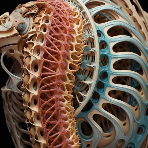
The human spinal cord, a key component of the central nervous system, is a long, thin, tubular structure made up of nervous tissue. It extends from the medulla oblongata in the brainstem to the lumbar region of the vertebral column.


The spinal cord is encased within the vertebral column and is further protected by three layers of tissue, known as meninges, and the cerebrospinal fluid. The cord is divided into 31 segments, each giving rise to a pair of spinal nerves. These segments are classified into four main regions: cervical, thoracic, lumbar, and sacral.
Function of the Spinal Cord
The spinal cord serves two primary functions. Firstly, it acts as a conduit for motor information, which travels down the spinal cord. Secondly, it acts as a conduit for sensory information, which travels up the spinal cord. This bidirectional flow of information is facilitated by the complex structure of the spinal cord, which is organized into grey and white matter.
Grey and White Matter
The spinal cord is composed of grey and white matter. The grey matter makes up the core of the spinal cord and is shaped like a butterfly or the letter "H". It contains cell bodies, dendrites and axon terminals, where synapses are found. The grey matter is responsible for processing and relaying information.
Surrounding the grey matter is the white matter, which is composed of bundles of myelinated axons, known as tracts. These tracts carry signals between the brain and the rest of the body. The white matter is divided into three funiculi: anterior, lateral, and posterior.
Spinal Cord Injury
Spinal cord injuries can result in a loss of motor function, sensation, or autonomic function in parts of the body served by the spinal cord below the level of the lesion. Injuries can occur at any level of the spinal cord and can be classified as complete injury, a total loss of sensation and muscle control, or incomplete, meaning some nervous signals are able to travel past the injured area of the cord.
