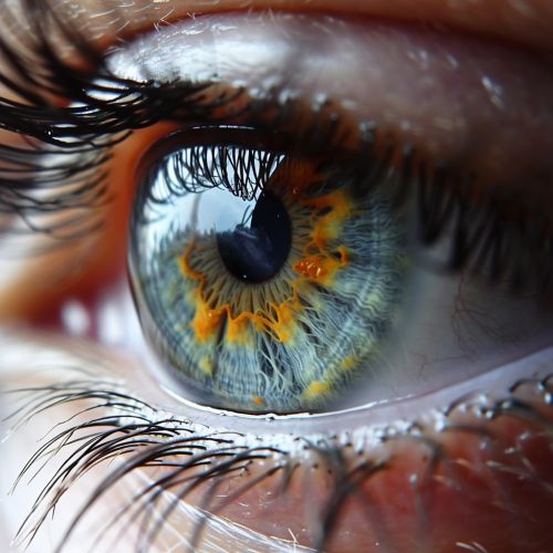
Dilator Pupillae
Anatomy and Physiology
The dilator pupillae is a muscle of the eye, specifically found in the iris. It is responsible for dilating or enlarging the size of the pupil in response to changes in light or during certain emotional states.


Structure
The dilator pupillae, also known as the radial muscle of the iris, is a thin layer of smooth muscle fibers. These fibers are arranged radially, extending from the outer edge of the iris to the inner edge of the pupil. This radial arrangement allows the muscle to effectively control the size of the pupil.
Function
The primary function of the dilator pupillae is to control the amount of light that enters the eye. It does this by adjusting the size of the pupil, the opening in the center of the iris through which light enters. When the dilator pupillae contracts, it pulls the iris outward, enlarging the pupil and allowing more light to enter. This is particularly useful in low-light conditions, where maximizing the amount of incoming light can improve vision.
Neural Control
The dilator pupillae is controlled by the sympathetic nervous system. This part of the nervous system is responsible for the body's 'fight or flight' response, and it triggers the dilation of the pupils in response to stress or excitement. This is known as mydriasis.
Clinical Significance
Abnormalities in the function of the dilator pupillae can lead to a variety of eye conditions. For example, damage to the sympathetic nerves that control the muscle can result in a condition known as Horner's syndrome, which is characterized by a constricted pupil, drooping eyelid, and loss of sweating on one side of the face.
