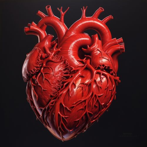
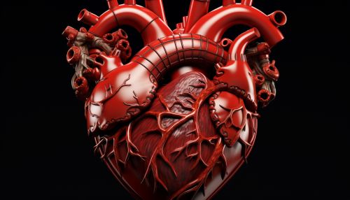
Anatomy of the heart
Anatomy of the Heart
The human heart, an organ of paramount importance in the circulatory system, is a complex structure with a variety of components, each with its own unique function and role. This article will delve into the intricate anatomy of the heart, providing a comprehensive and detailed examination of its structure, function, and the specialized terms associated with it.
Structure of the Heart
The heart is a muscular organ roughly the size of a closed fist, located slightly left of the center of the chest. It is divided into four chambers: the left atrium, the right atrium, the left ventricle, and the right ventricle.


Atria
The atria are the two upper chambers of the heart. The right atrium receives deoxygenated blood from the body through the superior and inferior vena cava and pumps it into the right ventricle. The left atrium, on the other hand, receives oxygenated blood from the lungs through the pulmonary veins and pumps it into the left ventricle.
Ventricles
The ventricles are the two lower chambers of the heart. The right ventricle receives deoxygenated blood from the right atrium and pumps it to the lungs through the pulmonary artery. The left ventricle, which is the strongest chamber, receives oxygenated blood from the left atrium and pumps it to the rest of the body through the aorta.
Heart Valves
The heart contains four valves: the tricuspid valve, the pulmonary valve, the mitral valve, and the aortic valve. These valves ensure unidirectional blood flow through the heart. The tricuspid valve is located between the right atrium and right ventricle, while the pulmonary valve is located between the right ventricle and pulmonary artery. The mitral valve, also known as the bicuspid valve, is located between the left atrium and left ventricle, and the aortic valve is located between the left ventricle and aorta.
Function of the Heart
The primary function of the heart is to pump blood throughout the body, supplying oxygen and nutrients to the tissues and removing carbon dioxide and other wastes. This is achieved through a process known as the cardiac cycle, which consists of two main phases: systole and diastole.
Systole
Systole is the phase of the cardiac cycle during which the ventricles contract, pumping blood out of the heart. During this phase, the aortic and pulmonary valves are open, allowing blood to flow into the aorta and pulmonary artery, respectively.
Diastole
Diastole is the phase of the cardiac cycle during which the ventricles relax and fill with blood. During this phase, the tricuspid and mitral valves are open, allowing blood to flow from the atria into the ventricles.
Specialized Terms
There are numerous specialized terms associated with the anatomy of the heart. These include:
- Myocardium: The muscular tissue of the heart.
- Endocardium: The inner lining of the heart.
- Epicardium: The outer layer of the heart wall.
- Pericardium: The sac that surrounds the heart.
- Coronary arteries: The vessels that supply blood to the heart muscle.
- Cardiac conduction system: The system of specialized cells and pathways that initiate and coordinate the contraction of the heart muscle.
