Anatomy of the Human Ear
Anatomy of the Human Ear
The human ear, an organ of hearing and balance, is a complex structure composed of three sections: the outer ear, the middle ear, and the inner ear. Each section performs distinct functions in the process of converting sound waves into electrical signals that the brain can interpret.
Outer Ear
The outer ear, also known as the auricle or pinna, is the visible part of the ear that resides outside of the head. It is primarily composed of cartilage, and its main function is to gather sound energy and direct it further into the ear. The outer ear also includes the ear canal, a tube running from the outer ear to the middle ear.
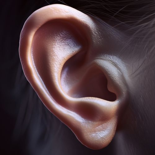
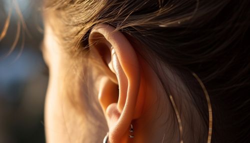
The auricle's shape is designed to capture sound waves and funnel them into the ear canal. The ridges and folds of the auricle can enhance certain frequencies of sound based on their size and shape. The ear canal, also known as the external auditory meatus, is a tube approximately 2.5 centimeters long in adults. It directs sound waves from the auricle to the tympanic membrane, or eardrum, in the middle ear.
Middle Ear
The middle ear is an air-filled cavity located behind the eardrum. It contains three tiny bones, collectively known as the ossicles, which are the smallest bones in the human body. These bones are named the malleus (hammer), incus (anvil), and stapes (stirrup) due to their distinctive shapes.
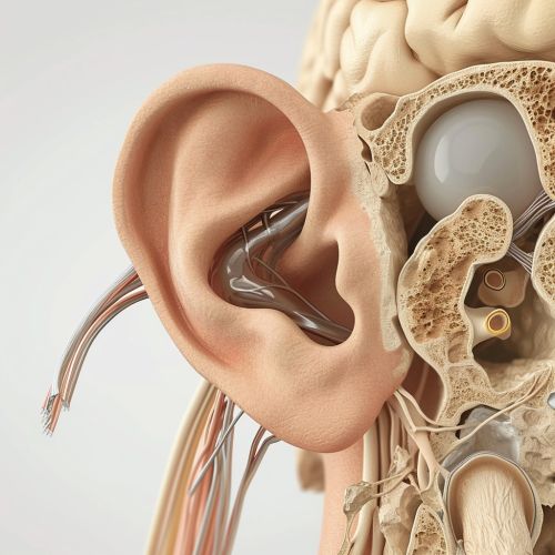
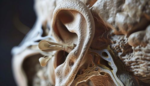
The eardrum, a thin, cone-shaped membrane, separates the outer ear from the middle ear. When sound waves reach the eardrum, they cause it to vibrate. These vibrations are then transferred to the ossicles. The ossicles amplify these vibrations and transmit them to the inner ear.
The middle ear also contains the Eustachian tube, which equalizes air pressure on both sides of the eardrum. This tube connects the middle ear to the nasopharynx, the upper part of the throat and the back of the nasal cavity.
Inner Ear
The inner ear, also known as the labyrinth, is the innermost part of the ear. It consists of two main structures: the cochlea, responsible for hearing, and the vestibular system, responsible for balance.
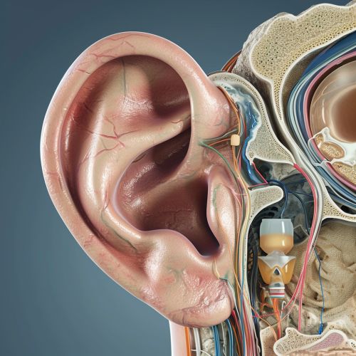
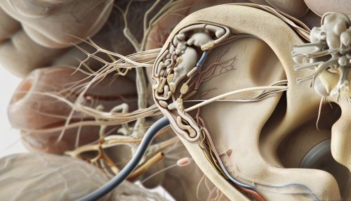
The cochlea is a spiral-shaped organ filled with fluid. When the ossicles in the middle ear transmit sound vibrations to the cochlea, the fluid inside it moves. This movement stimulates the hair cells inside the cochlea. These hair cells then convert the vibrations into electrical signals, which are sent to the brain via the auditory nerve.
The vestibular system consists of three semicircular canals and two otolith organs, the utricle and the saccule. The semicircular canals detect rotational movements of the head, while the otolith organs detect linear accelerations and gravity.
Disorders of the Ear
There are numerous disorders that can affect the human ear and its ability to hear and maintain balance. These include conductive hearing loss, sensorineural hearing loss, Meniere's disease, tinnitus, otitis media, and otosclerosis, among others. Each of these conditions can significantly impact a person's quality of life and may require medical intervention.
