Advances in Optical Coherence Tomography for Medical Imaging
Introduction
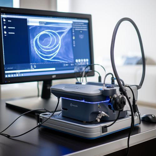
Optical Coherence Tomography (OCT) is a non-invasive imaging technique that uses light to capture micrometer-resolution, three-dimensional images from within optical scattering media such as biological tissue. It has been a significant advancement in the field of medical imaging, providing detailed, real-time visualizations of tissue structure and pathology. This article delves into the advancements in OCT technology, its applications, and future prospects.


History and Development
The concept of OCT was first introduced in the early 1990s, with the aim of improving the resolution and depth of imaging in biological tissue. The initial OCT systems were time-domain systems, where the depth information was obtained by mechanically varying the reference arm length in an interferometer. However, these systems were relatively slow and had limited imaging depth.
The introduction of Fourier-domain OCT (FD-OCT) in the early 2000s marked a significant advancement in OCT technology. FD-OCT, also known as spectral-domain OCT, uses a spectrometer in the detection arm of the interferometer to measure the interference spectrum. This allows for faster image acquisition and improved sensitivity compared to time-domain OCT.
In recent years, there has been a shift towards swept-source OCT (SS-OCT), which uses a tunable laser source to sweep the wavelength rapidly. SS-OCT offers even higher imaging speeds and deeper penetration into tissue, making it particularly useful for ophthalmic and cardiac imaging.
Technical Aspects
OCT operates on the principle of low-coherence interferometry. In simple terms, it measures the echo time delay and intensity of backscattered light from the tissue. The light source used in OCT is typically in the near-infrared range, as this wavelength allows for deeper penetration into tissue.
The axial resolution of OCT is determined by the coherence length of the light source, while the lateral resolution is determined by the focusing of the light beam. Modern OCT systems can achieve an axial resolution of a few micrometers, allowing for detailed visualization of tissue microstructure.
One of the key advantages of OCT is its ability to provide real-time, in vivo imaging. This makes it a valuable tool for guiding surgical procedures and monitoring disease progression.
Applications
OCT has found widespread application in various fields of medicine, particularly in ophthalmology and cardiology.
In ophthalmology, OCT is used to image the retina and anterior segment of the eye. It can detect subtle changes in retinal thickness and morphology, making it a powerful tool for diagnosing and monitoring diseases such as glaucoma, macular degeneration, and diabetic retinopathy.
In cardiology, OCT is used for intravascular imaging to assess coronary artery disease. It can provide detailed images of the vessel wall and plaque characteristics, aiding in the diagnosis and treatment planning of coronary artery disease.
Other applications of OCT include dermatology, gastroenterology, and oncology. In these fields, OCT is used for imaging skin lesions, gastrointestinal tract, and tumor margins, respectively.
Future Prospects
The field of OCT continues to evolve, with ongoing research aimed at improving image quality, speed, and depth of imaging. One promising area of research is the development of functional OCT techniques, which can provide additional information about tissue properties.
For example, Doppler OCT can measure blood flow velocity, while polarization-sensitive OCT can provide information about tissue birefringence. These techniques have the potential to enhance the diagnostic capabilities of OCT.
Another area of research is the integration of OCT with other imaging modalities, such as ultrasound or fluorescence imaging. This could provide complementary information and improve the diagnostic accuracy of OCT.
