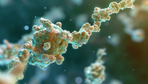
Tumor necrosis factor receptor
Introduction
Tumor necrosis factor receptor (TNFR) is a group of receptors that bind to tumor necrosis factors (TNFs). TNFs are a family of cytokines, which are proteins that mediate cell signaling and communication. The interaction between TNFs and their receptors plays a crucial role in numerous physiological and pathological processes, including cell survival, apoptosis, inflammation, and immune response.


Structure and Function
TNFRs are type I transmembrane proteins, meaning they span the cell membrane once and have their N-terminus on the outside of the cell. They are characterized by the presence of cysteine-rich domains (CRDs) in their extracellular region, which are responsible for binding to TNFs.
TNFRs can be activated by two types of TNFs: TNF-alpha and TNF-beta. TNF-alpha is primarily produced by macrophages, while TNF-beta is produced by lymphocytes. Upon binding to their respective receptors, these TNFs trigger a cascade of intracellular signaling events that can lead to various outcomes, depending on the cellular context.
Types of TNFRs
There are two main types of TNFRs: TNFR1 and TNFR2.
TNFR1
TNFR1, also known as p55 or CD120a, is expressed in most tissues and can be fully activated by both soluble and membrane-bound TNF. It contains a death domain (DD) in its intracellular region, which is essential for transmitting apoptotic signals.
TNFR2
TNFR2, also known as p75 or CD120b, is found primarily in cells of the immune system, such as T cells and macrophages. Unlike TNFR1, TNFR2 does not contain a DD and is not directly involved in apoptosis. Instead, it is primarily involved in promoting cell survival and proliferation.
Signaling Pathways
TNFRs can initiate several different signaling pathways, depending on the type of TNF they bind to and the cellular context.
Apoptosis
Apoptosis, or programmed cell death, is one of the primary responses triggered by TNFR1 activation. This process is initiated when the DD of TNFR1 recruits adapter proteins, such as TRADD and FADD, which in turn recruit and activate caspases, the enzymes responsible for executing apoptosis.
NF-kB Activation
Both TNFR1 and TNFR2 can activate the nuclear factor-kappa B (NF-kB) pathway, which plays a critical role in regulating immune response and inflammation. This pathway is triggered when TNFRs recruit adapter proteins, such as TRAF2 and RIP, which activate the IKK complex. The IKK complex then phosphorylates and degrades the inhibitor of NF-kB, allowing NF-kB to translocate to the nucleus and activate gene transcription.
Clinical Significance
Due to their role in regulating cell survival, apoptosis, and immune response, TNFRs are implicated in a variety of diseases, including autoimmune disorders, cancer, and infectious diseases.
Autoimmune Disorders
In autoimmune disorders, such as rheumatoid arthritis and systemic lupus erythematosus, TNFRs are thought to contribute to disease progression by promoting chronic inflammation and tissue damage.
Cancer
In cancer, TNFRs can have dual roles. On one hand, they can promote tumor growth and survival by activating the NF-kB pathway. On the other hand, they can induce apoptosis in cancer cells, thereby inhibiting tumor growth.
Infectious Diseases
In infectious diseases, TNFRs play a crucial role in coordinating the immune response against pathogens. However, excessive or prolonged activation of TNFRs can lead to tissue damage and disease.
