Sphincter Pupillae
Anatomy and Physiology
The Sphincter pupillae, also known as the pupillary sphincter, is a muscle in the human eye. It is a circular muscle that contracts to constrict the pupil in response to bright light or during the process of accommodation. This muscle is part of the autonomic nervous system and is innervated by the parasympathetic system.


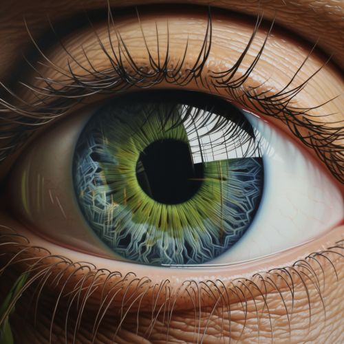
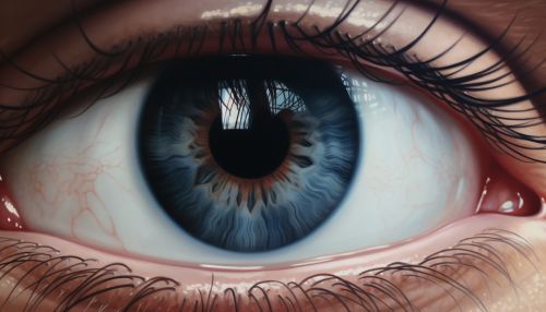
The sphincter pupillae is located in the iris, the colored part of the eye. It is found at the edge of the pupil and forms a ring around it. The muscle fibers are arranged in a circular pattern, allowing the muscle to contract in a way that reduces the size of the pupil.
Function
The primary function of the sphincter pupillae is to control the size of the pupil. It does this by contracting in response to bright light, a process known as the pupillary light reflex. This reflex helps to protect the retina from potential damage caused by excessive light exposure.
The sphincter pupillae also plays a role in the process of accommodation, which is the adjustment of the eye's optical power to maintain a clear image or focus on an object as its distance varies. During accommodation, the sphincter pupillae contracts to constrict the pupil, thereby increasing the depth of focus of the eye and reducing spherical aberration.
Innervation
The sphincter pupillae is innervated by the parasympathetic nervous system. The parasympathetic fibers that supply the sphincter pupillae originate from the Edinger-Westphal nucleus of the oculomotor nerve (cranial nerve III). These fibers travel with the oculomotor nerve and synapse in the ciliary ganglion, from which short ciliary nerves carry the parasympathetic fibers to the sphincter pupillae.
Clinical Significance
Abnormalities in the function of the sphincter pupillae can lead to a variety of clinical conditions. For example, damage to the oculomotor nerve can result in a dilated pupil (mydriasis) due to loss of parasympathetic innervation to the sphincter pupillae. This is often accompanied by other symptoms such as drooping of the eyelid (ptosis) and double vision (diplopia).
Another condition related to the sphincter pupillae is Adie's pupil, a neurological condition characterized by a dilated pupil that responds slowly to light but reacts normally to accommodation. This condition is thought to be caused by damage to the postganglionic fibers of the ciliary ganglion that supply the sphincter pupillae.
