SNARE
Overview
The Soluble NSF Attachment Protein Receptor (SNARE) proteins are a large protein complex that is the primary driver for membrane fusion in the cell, a critical process for intracellular trafficking. SNARE proteins are found in all eukaryotic cells and are essential for a variety of cellular processes, including vesicle fusion with the plasma membrane, vesicle fusion in the early stages of the secretory pathway, and autophagy.


Structure
SNARE proteins are characterized by a common SNARE motif, a stretch of 60-70 amino acids that can form a coiled-coil structure. This motif is found in all SNARE proteins and is essential for their function. The SNARE motif is usually located near the C-terminus of the protein and is followed by a transmembrane domain or a lipid anchor that tethers the protein to the membrane.
SNARE proteins can be classified into two categories based on the presence of a specific amino acid in the center of the SNARE motif: R-SNAREs, which contain an arginine (R) residue, and Q-SNAREs, which contain a glutamine (Q) residue. The R-SNAREs are often referred to as v-SNAREs because they are typically found on vesicles, while the Q-SNAREs are often referred to as t-SNAREs because they are typically found on target membranes.
Mechanism
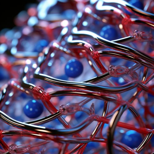
The SNARE proteins function by forming a SNARE complex, a four-helix bundle that brings the vesicle and target membranes into close proximity, allowing them to fuse. The formation of the SNARE complex is a highly exothermic reaction, providing the energy required for membrane fusion.
The SNARE complex is formed in a zippering process, starting from the N-terminus of the SNARE motif and progressing towards the C-terminus. This zippering process pulls the vesicle and target membranes together, overcoming the repulsive forces between the membranes and driving membrane fusion.
Once the fusion is complete, the SNARE complex is disassembled by the AAA+ ATPase NSF and its cofactor SNAP, allowing the SNARE proteins to be recycled for another round of fusion.
Role in Cellular Processes
SNARE proteins play a critical role in a variety of cellular processes, including exocytosis, endocytosis, and autophagy. In exocytosis, SNARE proteins mediate the fusion of secretory vesicles with the plasma membrane, allowing the release of the vesicle contents into the extracellular space. In endocytosis, SNARE proteins mediate the fusion of endocytic vesicles with early endosomes, allowing the vesicle contents to be sorted and recycled. In autophagy, SNARE proteins mediate the fusion of autophagosomes with lysosomes, allowing the degradation of the autophagosome contents.
SNARE proteins are also involved in the fusion of vesicles in the early stages of the secretory pathway, mediating the fusion of COPII-coated vesicles with the Golgi apparatus, and the fusion of COPI-coated vesicles with the endoplasmic reticulum.
Role in Disease
Mutations in SNARE proteins have been linked to a variety of diseases, including neurological disorders, immunodeficiencies, and cancer. For example, mutations in the gene encoding the R-SNARE protein VAMP2 have been associated with a form of familial epilepsy, while mutations in the gene encoding the Q-SNARE protein STX11 have been associated with a form of familial hemophagocytic lymphohistiocytosis, a severe immune disorder.
In addition, SNARE proteins have been implicated in the pathogenesis of several infectious diseases, as many pathogens have evolved mechanisms to manipulate the SNARE-mediated fusion machinery to facilitate their entry into host cells.
