Primary motor cortex
Anatomy and Location
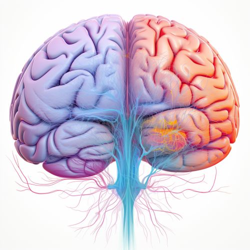
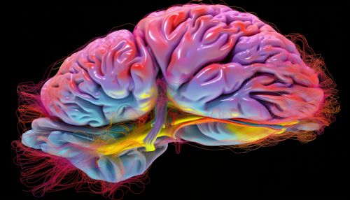
The primary motor cortex, also known as M1, is a section of the brain's cerebral cortex. It is located in the posterior portion of the frontal lobe, just before the central sulcus. The primary motor cortex is responsible for generating neural impulses that control the execution of movement. It is the most anterior (forward) part of the frontal lobe and is located on the precentral gyrus, which is the site of the primary motor area.


Function
The primary motor cortex plays a key role in the planning, control, and execution of voluntary movements. It is particularly involved in the control of precise and skilled movements, as well as the control of force. It is organized somatotopically, meaning that specific parts of the body are represented in specific parts of the primary motor cortex. This is often referred to as the motor homunculus, which is a physical representation of the body within the brain.
Neurophysiology
The neurons in the primary motor cortex send their axons down to the brainstem and spinal cord to synapse on motor neurons, which then connect to the muscles. The primary motor cortex contains large neurons known as Betz cells, which are a type of pyramidal neuron. These cells send long axons down the spinal cord to synapse on lower motor neurons in the spinal cord. The activity of the primary motor cortex is modulated by various other areas of the brain, including the premotor cortex, the supplementary motor area, and the basal ganglia, among others.
Clinical Significance
Damage to the primary motor cortex can result in a variety of motor deficits, including weakness or paralysis on the opposite side of the body (contralateral weakness or paralysis), loss of fine movements, and impairment of motor skills. These symptoms are often seen in conditions such as stroke, traumatic brain injury, and neurodegenerative diseases such as Amyotrophic lateral sclerosis (ALS) and Parkinson's disease.
Research and Future Directions
Research on the primary motor cortex has provided valuable insights into the neural control of movement. Techniques such as transcranial magnetic stimulation (TMS) and functional magnetic resonance imaging (fMRI) have been used to study the primary motor cortex in both healthy individuals and those with neurological disorders. Future research directions include the development of neuroprosthetic devices that can interface with the primary motor cortex to restore movement in individuals with paralysis.
