Muscular system
Overview
The muscular system is a complex network of tissues responsible for movement, stability, and various bodily functions. This system is composed of three types of muscle tissues: skeletal, cardiac, and smooth muscles. Each type of muscle tissue has distinct characteristics and functions, contributing to the overall operation of the body. The muscular system works in conjunction with the skeletal system, nervous system, and circulatory system to facilitate movement and maintain homeostasis.
Types of Muscle Tissue
Skeletal Muscle
Skeletal muscles are voluntary muscles attached to bones by tendons. They are responsible for skeletal movements such as walking, running, and lifting. These muscles are striated, meaning they have a banded appearance due to the arrangement of actin and myosin filaments. Skeletal muscles are controlled by the somatic nervous system, which allows for conscious control over movements.


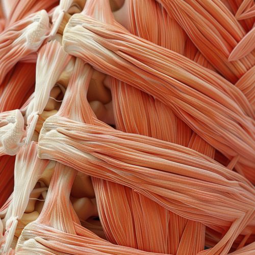
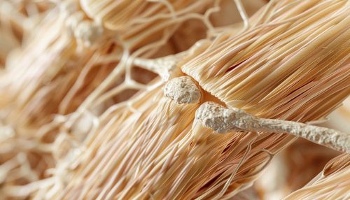
Skeletal muscles are composed of muscle fibers, which are long, cylindrical cells containing multiple nuclei. These fibers are bundled together by connective tissue to form a muscle. The functional unit of a skeletal muscle is the sarcomere, which is responsible for muscle contraction through the sliding filament theory.
Cardiac Muscle
Cardiac muscle is found exclusively in the heart. It is an involuntary, striated muscle that contracts rhythmically to pump blood throughout the body. Cardiac muscle cells, or cardiomyocytes, are branched and interconnected by intercalated discs, which facilitate synchronized contractions. These cells contain a single nucleus and are rich in mitochondria to meet the high energy demands of the heart.
Cardiac muscle contraction is regulated by the autonomic nervous system and intrinsic conduction system of the heart, including the sinoatrial node, atrioventricular node, and Purkinje fibers. This ensures the heart beats in a coordinated manner to maintain effective circulation.
Smooth Muscle
Smooth muscle is an involuntary, non-striated muscle found in the walls of hollow organs such as the intestines, blood vessels, and the bladder. Unlike skeletal and cardiac muscles, smooth muscle cells are spindle-shaped and contain a single nucleus. These muscles are responsible for various involuntary movements, including peristalsis in the digestive tract and vasoconstriction in blood vessels.
Smooth muscle contraction is regulated by the autonomic nervous system and various hormonal signals. The contraction mechanism involves the interaction of actin and myosin filaments, but the arrangement is less organized compared to striated muscles, resulting in a smooth appearance.
Muscle Contraction Mechanism
Muscle contraction is a complex process involving the interaction of actin and myosin filaments within the muscle fibers. This process is regulated by calcium ions and adenosine triphosphate (ATP). The sliding filament theory explains how muscles contract to produce force.
Sliding Filament Theory
The sliding filament theory describes the process by which muscle fibers contract. According to this theory, muscle contraction occurs when the thin actin filaments slide past the thick myosin filaments, shortening the sarcomere and thus the muscle fiber. This process is initiated by the binding of calcium ions to troponin, a regulatory protein on the actin filament. This binding causes a conformational change in tropomyosin, another regulatory protein, exposing the myosin-binding sites on actin.
Myosin heads then bind to these sites, forming cross-bridges. Using energy from ATP hydrolysis, the myosin heads pivot, pulling the actin filaments toward the center of the sarcomere. This action shortens the muscle fiber, generating force. The cycle repeats as long as calcium ions and ATP are present, resulting in sustained muscle contraction.
Muscle Metabolism
Muscle metabolism refers to the processes by which muscles generate energy for contraction and other functions. There are three primary pathways for ATP production in muscle cells: creatine phosphate pathway, anaerobic glycolysis, and aerobic respiration.
Creatine Phosphate Pathway
The creatine phosphate pathway provides a rapid source of ATP for short bursts of high-intensity activity. Creatine phosphate, stored in muscle cells, donates a phosphate group to adenosine diphosphate (ADP) to form ATP. This process is catalyzed by the enzyme creatine kinase and occurs within seconds, making it ideal for activities like sprinting or heavy lifting.
Anaerobic Glycolysis
Anaerobic glycolysis is a metabolic pathway that breaks down glucose to produce ATP in the absence of oxygen. This pathway generates ATP quickly but is less efficient than aerobic respiration, producing only two ATP molecules per glucose molecule. Anaerobic glycolysis also produces lactic acid as a byproduct, which can lead to muscle fatigue and soreness.
Aerobic Respiration
Aerobic respiration is the most efficient pathway for ATP production, generating up to 36 ATP molecules per glucose molecule. This process occurs in the mitochondria and requires oxygen. Aerobic respiration involves the complete oxidation of glucose through glycolysis, the citric acid cycle, and the electron transport chain. This pathway is essential for sustained, low-intensity activities such as long-distance running or cycling.
Muscle Adaptation and Plasticity
Muscle adaptation refers to the changes in muscle structure and function in response to various stimuli, such as exercise, injury, or disease. Muscle plasticity is the ability of muscle tissue to adapt to these changes, ensuring optimal performance and function.
Hypertrophy
Hypertrophy is the increase in muscle size due to an increase in the size of individual muscle fibers. This adaptation occurs in response to resistance training or other forms of high-intensity exercise. Hypertrophy involves the synthesis of new myofibrils and the addition of sarcomeres, resulting in increased muscle strength and power.
Atrophy
Atrophy is the decrease in muscle size due to a reduction in the size of individual muscle fibers. This condition can result from disuse, aging, or disease. Muscle atrophy involves the breakdown of myofibrils and the loss of sarcomeres, leading to decreased muscle strength and function.
Muscle Fiber Type Transformation
Muscle fibers can undergo transformation in response to different types of exercise. There are two main types of muscle fibers: slow-twitch (Type I) and fast-twitch (Type II). Slow-twitch fibers are more resistant to fatigue and are suited for endurance activities, while fast-twitch fibers generate more force and are suited for short bursts of high-intensity activity.
Endurance training can induce a shift from fast-twitch to slow-twitch fibers, enhancing the muscle's oxidative capacity and endurance. Conversely, resistance training can induce a shift from slow-twitch to fast-twitch fibers, increasing the muscle's strength and power.
Muscle Disorders and Diseases
The muscular system can be affected by various disorders and diseases, impacting muscle function and overall health. Some common muscle disorders include muscular dystrophy, myasthenia gravis, and rhabdomyolysis.
Muscular Dystrophy
Muscular dystrophy is a group of genetic disorders characterized by progressive muscle weakness and degeneration. The most common form is Duchenne muscular dystrophy, caused by mutations in the dystrophin gene. This disorder primarily affects boys and leads to severe disability and early death. Other forms of muscular dystrophy include Becker muscular dystrophy, myotonic dystrophy, and limb-girdle muscular dystrophy.
Myasthenia Gravis
Myasthenia gravis is an autoimmune disorder that affects the neuromuscular junction, leading to muscle weakness and fatigue. The immune system produces antibodies that block or destroy acetylcholine receptors on the muscle cell surface, preventing effective communication between nerves and muscles. Symptoms include ptosis, diplopia, and generalized muscle weakness. Treatment options include medications, plasmapheresis, and thymectomy.
Rhabdomyolysis
Rhabdomyolysis is a condition characterized by the rapid breakdown of muscle tissue, releasing myoglobin and other cellular contents into the bloodstream. This can lead to kidney damage and other complications. Causes of rhabdomyolysis include severe physical exertion, trauma, infections, and certain medications. Symptoms include muscle pain, weakness, and dark urine. Treatment involves addressing the underlying cause and providing supportive care to prevent kidney damage.
