Magnetic Resonance Force Microscopy
Introduction
Magnetic Resonance Force Microscopy (MRFM) is a type of scanning probe microscopy that measures the tiny magnetic forces between a magnetic tip and the magnetic spins in the sample. It is a highly sensitive tool that allows for the detection and manipulation of individual electron and nuclear spins, making it a powerful technique for studying a wide range of materials.
Principle of Operation
The basic principle of MRFM involves the interaction of a magnetic tip with the spins in the sample. The tip is attached to a micro-mechanical cantilever, which can be made to oscillate. When the tip is brought close to the sample, the magnetic spins in the sample cause a change in the cantilever's oscillation. This change can be detected and used to create an image of the sample's magnetic structure.


Components of MRFM
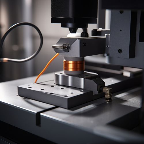
The main components of an MRFM setup include the cantilever with the magnetic tip, the sample, and the detection system. The cantilever is typically made from silicon or silicon nitride and is coated with a magnetic material to create the tip. The sample is placed on a stage that can be moved in all three dimensions, allowing for precise positioning of the sample relative to the tip. The detection system typically involves a laser and a photodiode to measure the cantilever's oscillation.
Cantilever and Magnetic Tip
The cantilever is a crucial component of the MRFM setup. It serves as the mechanical element that responds to the magnetic forces between the tip and the sample. The cantilever's oscillation is influenced by these forces, and this change in oscillation is what is ultimately detected and measured.
The magnetic tip at the end of the cantilever is typically made from a magnetic material such as iron or cobalt. The tip is magnetized, creating a magnetic field that interacts with the magnetic spins in the sample.
Sample Stage
The sample stage in an MRFM setup allows for precise positioning of the sample relative to the magnetic tip. The stage can be moved in all three dimensions (x, y, and z), allowing the tip to scan the sample in a raster pattern. This movement is typically controlled by piezoelectric actuators, which can provide very precise and controlled movement.
Detection System
The detection system in an MRFM setup is responsible for measuring the cantilever's oscillation. This is typically done using a laser and a photodiode. The laser is reflected off the back of the cantilever, and the reflected light is detected by the photodiode. Any change in the cantilever's oscillation will cause a change in the position of the reflected laser light, which can be detected by the photodiode.
Applications of MRFM
MRFM has a wide range of applications in various fields including physics, chemistry, and biology. It can be used to study the magnetic properties of materials, to investigate the structure of molecules, and to image biological samples at the nanoscale.
Physics
In physics, MRFM can be used to study the magnetic properties of materials. For example, it can be used to investigate the magnetic domains in ferromagnetic materials or to study the spin dynamics in magnetic systems. MRFM can also be used to investigate quantum phenomena, such as quantum entanglement and quantum superposition, at the nanoscale.
Chemistry
In chemistry, MRFM can be used to investigate the structure of molecules. By measuring the magnetic interactions between the magnetic tip and the spins in the sample, MRFM can provide information about the spatial arrangement of atoms in a molecule. This can be particularly useful in the study of complex organic molecules and biomolecules.
Biology
In biology, MRFM can be used to image biological samples at the nanoscale. For example, it can be used to study the structure of proteins or to investigate the organization of cellular components. MRFM can also be used to study the magnetic properties of biological systems, such as the magnetic sensing abilities of certain organisms.
Advantages and Limitations
Like all scientific techniques, MRFM has its advantages and limitations. One of the main advantages of MRFM is its high sensitivity. It can detect and manipulate individual electron and nuclear spins, making it a powerful tool for studying a wide range of materials. MRFM also has a high spatial resolution, allowing for the imaging of samples at the nanoscale.
However, MRFM also has some limitations. The technique requires a high level of precision and control, both in the preparation of the sample and in the operation of the MRFM setup. Additionally, the interpretation of MRFM data can be complex, requiring a deep understanding of magnetic resonance and spin dynamics.
