Light Microscopy
Introduction
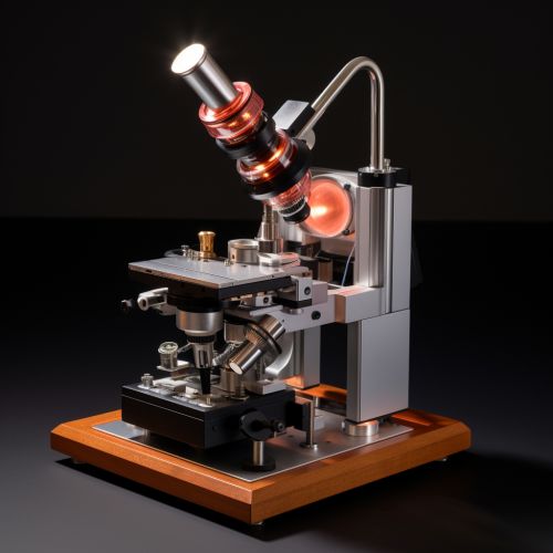
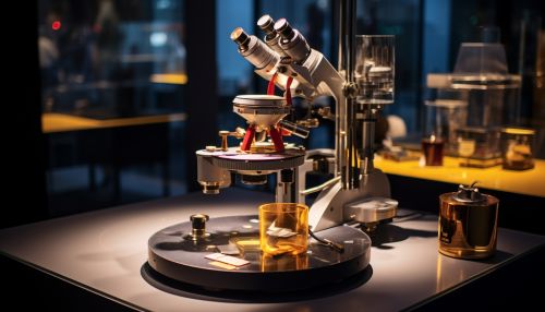
Light microscopy is a technique that utilizes visible light and a system of lenses to magnify images of small samples. First developed in the 17th century, it has since been refined and expanded upon, leading to a variety of specialized light microscopes with different imaging techniques and applications.


History
The first simple microscopes, which used a single lens to magnify the sample, were developed in the 16th and 17th centuries. The compound microscope, which uses multiple lenses to achieve higher magnification, was developed in the 17th century by Robert Hooke and Antonie van Leeuwenhoek. These early microscopes had a number of limitations, including low resolution and chromatic aberration, but they were still able to reveal a previously unseen world of microscopic organisms and structures.
Principles of Light Microscopy
The basic principle of light microscopy is the refraction, or bending, of light as it passes through a lens. When light passes from one medium (such as air) into another medium with a different refractive index (such as glass), it changes direction. This change in direction allows a lens to focus light onto a specific point, creating a magnified image of the sample.
The resolution of a microscope, or its ability to distinguish between two points that are close together, is determined by the wavelength of the light used and the numerical aperture of the objective lens. The numerical aperture is a measure of the lens's ability to gather light and resolve fine specimen detail at a fixed object distance.
Types of Light Microscopy
There are several different types of light microscopy, each with its own advantages and disadvantages. These include bright field microscopy, dark field microscopy, phase contrast microscopy, differential interference contrast (DIC) microscopy, fluorescence microscopy, and confocal microscopy.
Bright Field Microscopy
Bright field microscopy is the most common type of light microscopy. It involves light being passed directly through the sample, with any contrast in the image being caused by absorption of light by the sample. This technique is simple and easy to use, but it can lack contrast and detail when used with unstained or transparent samples.
Dark Field Microscopy
Dark field microscopy uses a special condenser to scatter light so that the sample appears bright against a dark background. This technique is useful for viewing transparent samples that do not absorb light well, such as living cells or organisms in water.
Phase Contrast Microscopy
Phase contrast microscopy converts phase shifts in light passing through a transparent specimen to brightness changes in the image. It is used to enhance the contrast in unstained, transparent samples.
Differential Interference Contrast (DIC) Microscopy
DIC microscopy uses polarized light to give a three-dimensional appearance to flat, transparent samples. It is particularly useful for examining the surfaces of cells and other biological specimens.
Fluorescence Microscopy
Fluorescence microscopy uses a specific wavelength of light to excite fluorescent dyes or proteins in the sample, causing them to emit light of a different wavelength. This technique is widely used in biological and medical research to study the distribution of proteins, nucleic acids, and other molecules within cells and tissues.
Confocal Microscopy
Confocal microscopy uses a laser to scan the sample point by point, creating a digital image that can be viewed and manipulated on a computer. This technique allows for the imaging of thick samples and the reconstruction of three-dimensional structures.
Applications of Light Microscopy
Light microscopy has a wide range of applications in various fields, including biology, medicine, materials science, and geology. In biology and medicine, it is used to study cells and tissues, diagnose diseases, and conduct research. In materials science, it is used to examine the structure and properties of various materials. In geology, it is used to study rocks and minerals.
